. 2023 Sep 26;120(39):e2300416120.
doi: 10.1073/pnas.2300416120. Epub 2023 Sep 19.

Friction patterns guide actin network contraction

Affiliations

Friction patterns guide actin network contraction

Alexandra Colin et al. Proc Natl Acad Sci U S A. .

Abstract

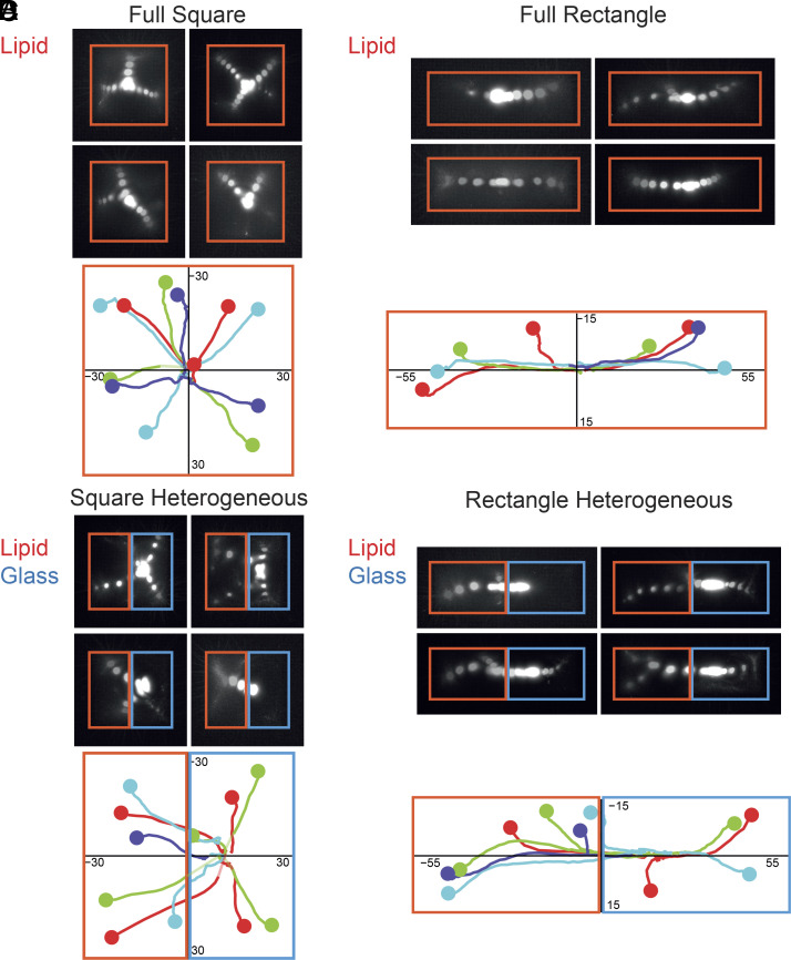
The shape of cells is the outcome of the balance of inner forces produced by the actomyosin network and the resistive forces produced by cell adhesion to their environment. The specific contributions of contractile, anchoring and friction forces to network deformation rate and orientation are difficult to disentangle in living cells where they influence each other. Here, we reconstituted contractile actomyosin networks in vitro to study specifically the role of the friction forces between the network and its anchoring substrate. To modulate the magnitude and spatial distribution of friction forces, we used glass or lipids surface micropatterning to control the initial shape of the network. We adapted the concentration of Nucleating Promoting Factor on each surface to induce the assembly of actin networks of similar densities and compare the deformation of the network toward the centroid of the pattern shape upon myosin-induced contraction. We found that actin network deformation was faster and more coordinated on lipid bilayers than on glass, showing the resistance of friction to network contraction. To further study the role of the spatial distribution of these friction forces, we designed heterogeneous micropatterns made of glass and lipids. The deformation upon contraction was no longer symmetric but biased toward the region of higher friction. Furthermore, we showed that the pattern of friction could robustly drive network contraction and dominate the contribution of asymmetric distributions of myosins. Therefore, we demonstrate that during contraction, both the active and resistive forces are essential to direct the actin network deformation.

Keywords: actin; contraction; cytoskeleton; friction.


Conflict of interest statement

The authors declare no competing interest.

Figures

Fig. 1.
Fig. 1.
Actin assembly on glass or lipid micropatterns. (A) Cartoon of the method to constrain branched actin network assembly on glass or lipid micropatterns. (B) TIRF imaging of branched actin assembly on lipid micropattern (disk, diameter 135 μm). Biochemical conditions: WA 1 nM, Actin 1 μM, Human Profilin 3 μM, Arp2/3 complex 25 nM. (Scale bar: 10 μm.) (C) Characterization of the diffusion property of the lipid micropattern. Left: TIRF imaging before and after FRAP (zone diameter: 10 μm) on lipids, NPF (WA), and Actin (after network polymerization). Biochemical conditions: disk micropattern (diameter 68 μm), WA 1 nM, Actin 1 μM, Human Profilin 3 μM, and Arp2/3 complex 25 nM. (The scale bar is 20 μm.) Right: Fluorescence measurements from the images on the left demonstrate that the lipids and the NPF diffuse freely in our experimental conditions. (D) Comparison of the efficiency of actin network growth on lipid versus glass micropatterns. TIRF imaging of branched actin assembly on lipid and glass disk micropatterns (diameter 68 μm). Biochemical conditions: NPF concentration is indicated on the figure. Actin 1 μM, Human Profilin 3 μM, and Arp2/3 complex 25 nM. (The scale bar is 20 μm.) (E) Kinetics of actin assembly on lipid versus glass micropattern.
Fig. 2.
Fig. 2.
NPF attachment conditions control actin network contractile response. (A) Kinetics of contraction of a disk-shaped actin network on glass or lipid micropattern. Time-lapse imaging of actin network contraction on a glass or lipid disk (diameter 68 μm) micropattern. The line (blue for glass; red for lipids) corresponds to the contours of the actin network (Materials and Methods). Biochemical conditions: On glass micropattern, WA = 1 μM; on lipid micropattern: WA = 1 nM. Actin 1 μM, Human Profilin 3 μM, Arp2/3 complex 25 nM, and Myosin VI 14 nM. (The scale bar is 20 μm.) (B) Kinetics of contraction of a square-shaped actin network on glass or lipid micropattern. Time-lapse imaging of actin network contraction on a glass or lipid square (length 60 μm) micropattern. The line (blue for glass; red for lipids) corresponds to the contours of the actin network (Materials and Methods). Biochemical conditions: On glass micropattern, WA = 1 μM; on lipid micropattern: WA = 1 nM. Actin 1 μM, Human Profilin 3 μM, Arp2/3 complex 25 nM, and Myosin VI 14 nM. (The scale bar is 20 μm.) (C and D) Measured actin area as a function of time for the lipid (red) or glass (red) conditions on disk (C) or square (D) micropattern. The period defined at the top of the graph determines the lag phase for each condition. (E) Duration of the lag phase preceding the contraction for the lipid or glass conditions on disk or square micropattern. Data are represented with a superplot. Disk glass: n = 40; N = 2; median = 7.5. Disk lipids n = 41; N = 3; median = 4.0. Square glass n = 37; N = 2; median = 7.0. Square lipids n = 37; N = 2; median = 4.0. Mann–Whitney statistics: disk glass/disk lipids P value = 0.0010***, square glass/square lipids P value ≤ 0.0001****, disk glass/square glass P value = 0.1247 ns, and disk lipids/square lipids P value = 0.5360 ns. (F) Velocity of the phase contraction phase for the lipid or glass conditions on disk or square micropattern. Data are represented with a superplot. Disk glass n = 53; N = 3; median = 0.8346, disk lipids n = 68; N = 4; median = 1.133, square glass n = 37; N=2; median = 1.013, and square lipids n = 37; N = 2; median = 1.694. Mann–Whitney statistics: disk glass/disk lipids P value ≤ 0.0001****, square glass/square lipids P value ≤ 0.0001****, disk glass/square glass P value = 0.2761 ns, and disk lipids/square lipids P value ≤ 0.0001****.
Fig. 3.
Fig. 3.
Lower friction improves the coordination of the contraction process. (A) Local deformation of the actin network. A photobleached grid shape was performed on actin networks polymerized on glass or lipid micropatterns. Then, deformations of the actin network were followed by following the grid deformation. (B) Left Top: Example of an actin network grown on glass substrate pattern used for PIV analysis. Left Bottom: PIV analysis of the actin network shown above. Right: Resultant of vector sum for each quadrant defined on the pattern as a function of time (Materials and Methods). (C) Same as B with an actin network grown on lipid substrate. (D) Left: Snapshots of the final position of myosin spot for actin networks grown on glass or lipid square micropattern. Right: Quantification of the distance between the final myosin spot and the pattern center for networks grown on glass or lipids. N = 2 independent replicates with n = 18 and n = 12 patterns for the lipids condition and n = 19 and n = 13 patterns for the glass condition. Individual points for each pattern are represented. Mean and SD are plotted on top of the points. Unpaired t test: P value ≤ 0.001***. Biochemical conditions for Fig. 3: On glass micropattern, WA = 1 μM; on lipid micropattern: WA = 1 nM. Actin 1 μM, Human Profilin 3 μM, Arp2/3 complex 25 nM, and Myosin VI 14 nM.
Fig. 4.
Fig. 4.
Friction pattern directs network contraction. Time-lapse imaging of actin network contraction on full lipid or heterogeneous disks (A), full lipid or heterogeneous squares (C), and full lipid or heterogeneous rectangles (E). Biochemical conditions: Actin 1 μM, Human Profilin 3 μM, Arp2/3 complex 25 nM, and Myosin VI 10 nM. Pattern dimensions: disks 64 µm diameter; squares 60 µm length; rectangles 104 µm × 34 µm. The pattern is represented in blue, actin in red, and myosin in green. (Scale bar: 20 μm.) (B, D, and F) Plot of the myosin dots coordinates at the end of contraction for the different shapes. All coordinates were measured and compared to the centroid (x = 0, y = 0) of the whole pattern. Red dots correspond to the coordinates of the full pattern made of lipid, green dots correspond to the coordinates of the symmetrical heterogeneous pattern and the blue dots correspond to the coordinates of the asymmetrical heterogeneous patterns. Disk full lipids: N = 3, n = 11 patterns. Heterogeneous disk: N = 4, n = 26 patterns. Square full lipids: N = 2, n = 9 patterns. Heterogeneous square: N = 4, n = 29 patterns. Asymmetrical heterogeneous square: N = 2, n = 11 patterns. Rectangle full lipids: N = 2, n = 8 patterns. Heterogeneous rectangle: N = 4, n = 24 patterns. Asymmetrical heterogeneous rectangle: N = 3, n = 21 patterns.
Fig. 5.
Fig. 5.
The computational model recapitulates the contraction kinetics and predicts the trajectories of myosin spots during the network contraction. (A) Scheme of the computational model describing the network as a viscoelastic network of nodes and links. Each node is connected by a dashpot with the substrate. The dashpot is responsible for the friction (higher on glass, lower on lipid). Neighboring nodes are connected by dashpot/spring in series responsible for viscous and elastic deformations inside the network. In addition, links connecting pairs of nodes, one of which is occupied by a myosin dot (shown by a star), are contractile. (B) Sequence of deformations predicted by the model for an actin network polymerized on a homogeneous square lipid micropattern and contracted by myosin dots (yellow nodes). (C) Prediction of myosin trajectories for an actin network contracting on a homogeneous square lipid micropattern. (D) Sequence of deformations predicted by the model for an actin network polymerized on a heterogeneous square micropattern and contracted by myosin dots (yellow nodes). (E) Prediction of myosin trajectories for an actin network contracting on a heterogeneous square micropattern.
Fig. 6.
Fig. 6.
Friction pattern robustly drives network contraction despite uneven distribution of myosin. Top: Four examples of temporal projections of myosin detection on a full-square micropattern (A), a full-rectangle micropattern (B), a heterogeneous square micropattern (C), and a heterogeneous rectangle micropattern (D). Bottom: Detection of myosin spots as a function of time (see Materials and Methods for details). Each example of the above images is represented with a single color. The initial time of the trajectory is represented with a bigger dot.

References

    1. Kollmar M., Mühlhausen S., Myosin repertoire expansion coincides with eukaryotic diversification in the Mesoproterozoic era. BMC Evol. Biol. 17, 211 (2017). - PMC - PubMed
    1. Hartman M. A., Spudich J. A., The myosin superfamily at a glance. J. Cell Sci. 125, 1627–1632 (2012). - PMC - PubMed
    1. Wickstead B., Gull K., The evolution of the cytoskeleton. J. Cell Biol. 194, 513–525 (2011). - PMC - PubMed
    1. Sellers J. R., Myosins: A diverse superfamily. Biochim. Biophys. Acta–Mol. Cell Res. 1499, 3–22 (2000). - PubMed
    1. Robert-Paganin J., Pylypenko O., Kikuti C., Sweeney H. L., Houdusse A., Force generation by myosin motors: A structural perspective. Chem. Rev. 120, 5–35 (2020). - PubMed

Publication types

LinkOut - more resources