. 2023 Sep 26;120(39):e2309955120.
doi: 10.1073/pnas.2309955120. Epub 2023 Sep 19.

Disassembly of bundled F-actin and cellular remodeling via an interplay of Mical, cofilin, and F-actin crosslinkers

Affiliations

Disassembly of bundled F-actin and cellular remodeling via an interplay of Mical, cofilin, and F-actin crosslinkers

Sudeepa Rajan et al. Proc Natl Acad Sci U S A. .

Abstract

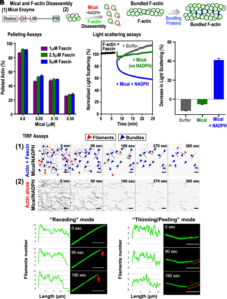
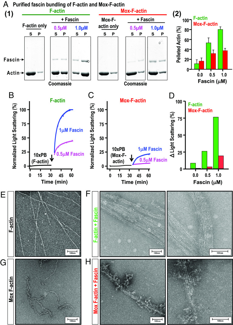
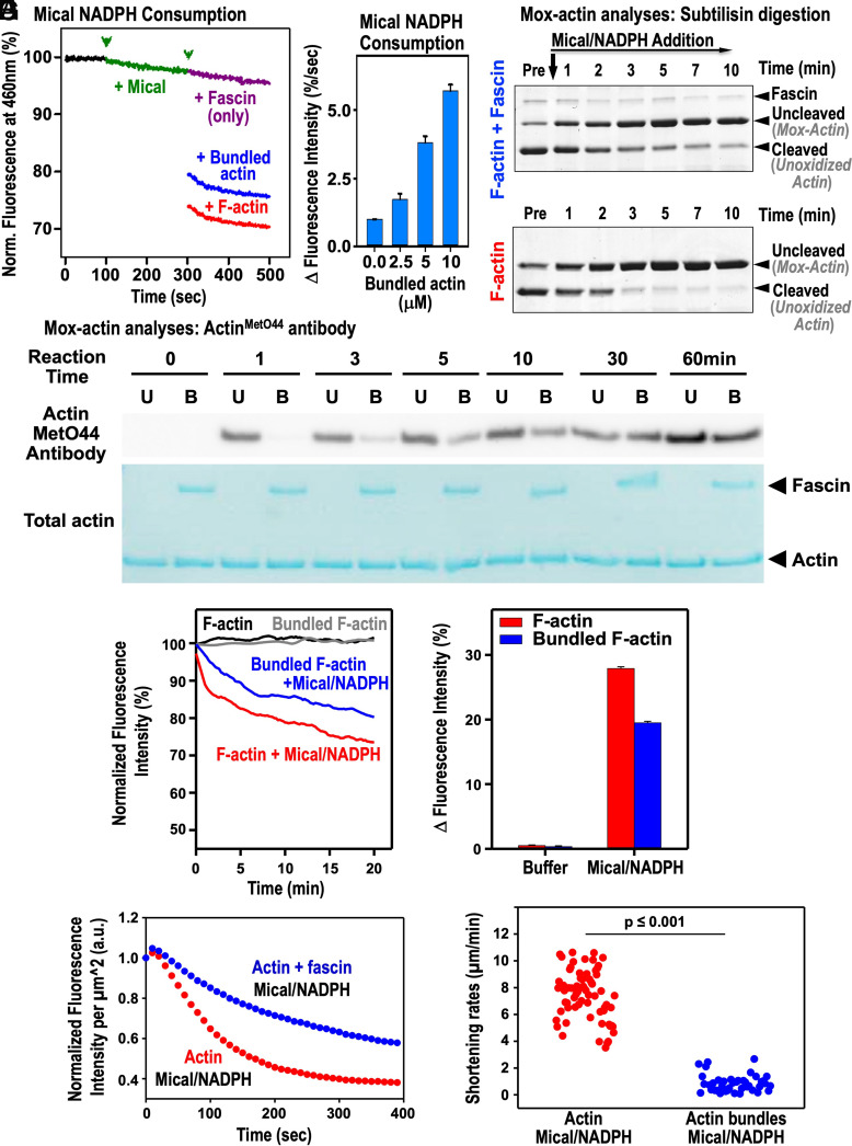
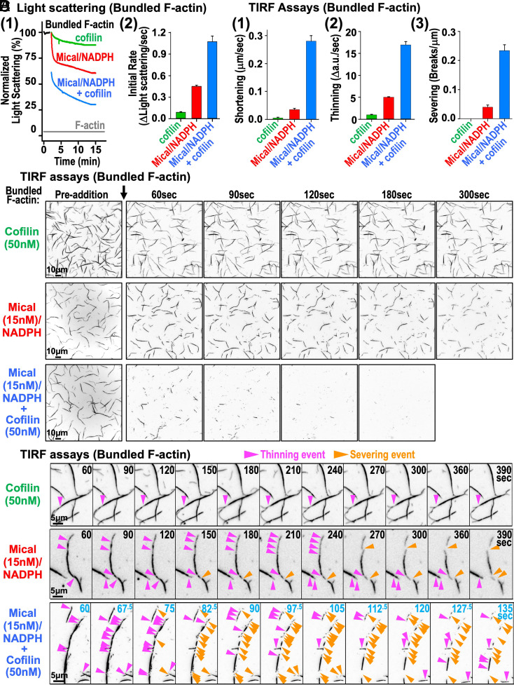
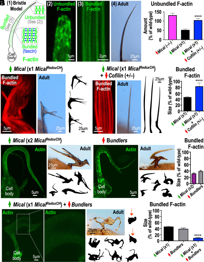
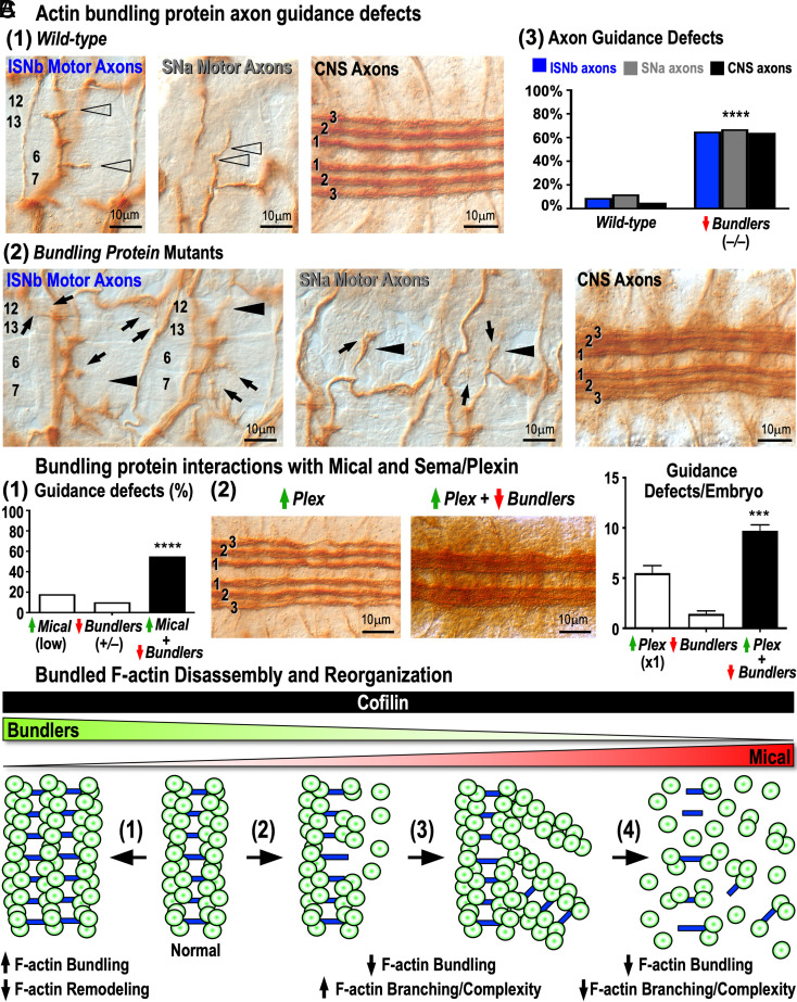
Cellular form and function are controlled by the assembly and stability of actin cytoskeletal structures-but disassembling/pruning these structures is equally essential for the plasticity and remodeling that underlie behavioral adaptations. Importantly, the mechanisms of actin assembly have been well-defined-including that it is driven by actin's polymerization into filaments (F-actin) and then often bundling by crosslinking proteins into stable higher-order structures. In contrast, it remains less clear how these stable bundled F-actin structures are rapidly disassembled. We now uncover mechanisms that rapidly and extensively disassemble bundled F-actin. Using biochemical, structural, and imaging assays with purified proteins, we show that F-actin bundled with one of the most prominent crosslinkers, fascin, is extensively disassembled by Mical, the F-actin disassembly enzyme. Furthermore, the product of this Mical effect, Mical-oxidized actin, is poorly bundled by fascin, thereby further amplifying Mical's disassembly effects on bundled F-actin. Moreover, another critical F-actin regulator, cofilin, also affects fascin-bundled filaments, but we find herein that it synergizes with Mical to dramatically amplify its disassembly of bundled F-actin compared to the sum of their individual effects. Genetic and high-resolution cellular assays reveal that Mical also counteracts crosslinking proteins/bundled F-actin in vivo to control cellular extension, axon guidance, and Semaphorin/Plexin cell-cell repulsion. Yet, our results also support the idea that fascin-bundling serves to dampen Mical's F-actin disassembly in vitro and in vivo-and that physiologically relevant cellular remodeling requires a fine-tuned interplay between the factors that build bundled F-actin networks and those that disassemble them.

Keywords: MICAL1; MICAL2; MICAL3; bristle; nervous system.


Conflict of interest statement

The authors declare no competing interest.

Figures

Fig. 1.
Fig. 1.
Mical robustly disassembles fascin-bundled F-actin. (A) Mical and its effects on F-actin. (1) Mical enzyme. Redox enzymatic, CH (calponin homology), LIM (Lin11, Isl-1, and Mec-3), and PIR (Plexin-interacting region) domains. (2) Mical in the presence of its coenzyme NADPH disassembles F-actin (green) by oxidizing (red O) it. (B) Actin bundling/crosslinking proteins bundle actin together in different organizations, such as parallel-arranged filaments. (C) Pelleting assays show that Mical disassembles fascin-bundled F-actin at different saturating concentrations of fascin and in a Mical concentration-dependent manner. See SI Appendix, Fig. S1D for gels. [F-actin] = 5 μM, [NADPH] = 400 µM. [Mical], [fascin] as indicated. n = 3 independent experiments/condition. Mean ± SEM. (D) Light scattering assays show that Mical/NADPH disassembles fascin-bundled F-actin. Normalized light (325 nm) scattering percentage (%) changes [also for Figs. 2 B and C and 4A (1) and SI Appendix, Figs. S1 E and F, S2D, and S3I]. Specifically, F-actin was bundled with fascin to a steady state (black), and then buffer with NADPH (gray), Mical (green), or Mical with NADPH (blue) was added to it. [F-actin] = 5 μM, [fascin] = 5 μM, [Mical] = 0.05 μM, and [NADPH] = 100 μM. A representative experiment is shown (Left). n = 3 independent experiments/condition. Mean ± SEM. Minutes (min). (E) F-actin disassembly with (1) or without (2) fascin upon on-slide Mical/NADPH oxidation. Representative TIRFM movie montages; bar = 10 µm. Red and blue arrowheads indicate unbundled and bundled filaments, respectively. Seconds (sec). (F and G) Predominant modes of Mical-mediated bundled-F-actin disassembly observed by TIRFM. (Left) fluorescence intensity profiles of bundles from indicated time points and corrected for photobleaching: average fluorescence intensities of unbundled filaments from same movies (internal controls) were used to determine filaments number/bundle (Y-axes). (Right) representative TIRFM movie montages; bar = 5 µm. (F) “Receding mode” is defined as bundle shortening (red arrow) without changing its maximal thickness. (G) “Thinning/Peeling mode” is defined as bundle shortening accompanied by its thinning (usually from one end). Red bracket = thinning region. Note that bundle ends can alternate between “receding” and “thinning/peeling” disassembly modes. [actin] = 1.24 µM; [fascin] = 0.8 µM; [Mical] = 10 nM; [NADPH] = 100 µM.
Fig. 2.
Fig. 2.
Fascin poorly bundles Mical-oxidized F-actin. (A) Low-speed pelleting. (1) Supernatant (S) and pellet (P) contents of F-actin and Mox-F-actin incubated without or with fascin. (2) Pelleted F-actin and Mox-F-actin percentage at different [fascin]. [actins] = 5 μM, [fascin] = see figure. Representative experiments are shown. n = 3 independent experiments/condition. Mean ± SEM. (BD) Light scattering assays. Black trace = actin polymerization using a 10×-concentrated polymerization buffer (PB). Black arrow=fascin addition. [fascin] = see figure. Representative experiments are shown. n = 3 independent experiments/condition. (EH) Transmission EM images of negatively stained F-actin alone (E), F-actin with fascin (F), Mox-F-actin alone (G), and Mox-F-actin with fascin (H) reveal that unmodified F-actin bundles are smooth and coherent while Mox-F-actin forms short, coarse, and disordered bundles. (F and H) low (Left) and high (Right) image magnification.
Fig. 3.
Fig. 3.
Fascin-bundling regulates Mical’s oxidation-mediated F-actin disassembly activity. (A and B) Mical’s enzymatic activity, as judged by NADPH consumption [conversion of NADPH to NADP+ (fluorescence decrease/change)] is activated by fascin-bundled F-actin but to a lesser extent than by unbundled F-actin. A = [F-actin] = 2.5 μM, [fascin] = 0.5 μM, [Mical] = 0.6 μM, [NADPH] = 200 μM. B = Mical’s increasing enzymatic activity with increasing [fascin-bundled F-actin]. n = 3 independent experiments/condition, mean ± SEM. (C and D) Mical oxidizes F-actin bundled by fascin, but at a slower rate than unbundled F-actin. (C) Subtilisin does not cleave Mical-oxidized actin between its M47 and G48 residues (24, 25). (Upper) Mical/NADPH’s addition decreases subtilisin’s cleavage of fascin-bundled F-actin over time. This reveals Mical/NADPH oxidizes fascin-bundled F-actin. (Lower) Mical/NADPH’s addition more rapidly decreases subtilisin’s cleavage of unbundled versus bundled F-actin (compare Cleaved Actin, Lower and Upper). This reveals fascin’s presence dampens Mical-mediated F-actin oxidation. Pre = prior to Mical/NADPH addition (note that a small amount of actin is not cleaved under our conditions even without Mical/NADPH present). [F-actin] = 3.5 μM, [fascin] = 0.7 µM, [Mical] = 0.01 µM, and [NADPH] = 100 μM. (D) ActinMetO44-specific antibody shows that Mical oxidizes fascin-bundled F-actin (B lanes), but this oxidation is slower than that seen with unbundled F-actin (U lanes). [F-actin] = 1.15 µM, [fascin] = 0.5 µM, [Mical] = 0.05 µM, and [NADPH] = 100 µM. (EG) Fascin-bundling slows Mical’s F-actin disassembly. (E) Pyrene-actin fluorescence (407 nm) assay. [F-actin] = 2.5 µM, [cofilin] = 0.25 µM, [Mical] = 0.025 µM, and [NADPH] = 100 µM. Representative experiment, n = 3 independent experiments/condition, mean ± SEM. (F) Average fluorescence intensities/µm2 for TIRFM movies reveal Mical/NADPH induces a ~twofold slower fluorescence signal decay in fascin-bundled F-actin (blue) compared to unbundled filaments (red). Data averaged from four movies. n = 3 separate experiments. (G) Shortening rate measurements show a statistically significant (8.5-fold) inhibition of Mical-mediated disassembly by fascin-bundled F-actin (n = 41, blue) compared to unbundled filaments (n = 66, red). Mann–Whitney test. Measurements from three separate experiments. [actin] = 1.24 µM, [fascin] = 0.8 µM, [Mical] = 10 nM, and [NADPH] = 100 µM.
Fig. 4.
Fig. 4.
Mical synergizes with cofilin to enhance fascin-bundled F-actin disassembly. Light scattering (A) and TIRF assays (BD) reveal that together Mical and cofilin disassemble bundled F-actin more rapidly and more effectively than the sum of separate cofilin and Mical disassembly rates. (A) Light scattering: Graphs show the changes (1) and the initial rates of change (2). [actin] = 5 μM; [fascin] = 1 μM; [cofilin] = 1 μM; [Mical] = 0.05 μM; and [NADPH] = 100 μM. A representative experiment is shown; n = 5. Mean ± SEM. (B) TIRF assays: graphs show the rates of bundles shortening (1), thinning (2), and severing (3) from experiments described in C and D (data are averaged from five different movies). n = 40, 27, and 35 bundles used for Mical, cofilin, and both Mical and cofilin conditions, respectively. Mean ± SEM. (C and D) Representative time-lapse TIRFM video montages of fascin-bundled F-actin disassembly in the presence of cofilin, Mical, and Mical + cofilin (arrow = addition of each). Note the shortened time frame in Mical + cofilin in D. [actin] = 1 μM, 20% Alexa labeled; [fascin] = 1 μM; [cofilin] = 50 nM; [Mical] = 15 nM; and [NADPH] = 100 μM.
Fig. 5.
Fig. 5.
Mical combines with cofilin to decrease bundled F-actin in vivo, but bundling proteins regulate this ability. (A) Bristle actin filaments (green) exist as both unbundled snarls/patches ((1) upper boxed region, (2)) and bundled F-actin ((1) lower boxed regions, (3)) crosslinked with bundling proteins (blue) to “push-out” long, branchless bristles (1 and 4). (B) Decreasing Mical in vivo increases unbundled F-actin and increasing Mical in vivo decreases unbundled F-actin in a cofilin-dependent manner. See SI Appendix, Fig. S4A for images. (CE) Increasing Mical (C and E) in a cofilin-dependent manner (D and E) decreases bundled F-actin in bristles in vivo. (FH) Increasing Mical to high levels (x2) in vivo (F and H) mimics the cellular and F-actin effects of decreasing bundling proteins in vivo (G and H). (I and J) Increasing Mical (x1) in combination with decreasing bundling proteins enhances the in vivo loss of bundled F-actin (green in I, and compare to altering either effector on its own in J) and generates bristles that have a rounded, cell-body-like shape (Adult, e.g., arrows). The dashed rectangular region is magnified in the adjacent image. Mean ± SEM for graphs, ****P < 0.0001, unpaired t test (two tailed) for B, E, and J.
Fig. 6.
Fig. 6.
Semaphorin/Plexin/Mical repulsion counteracts actin-bundling proteins to direct axon guidance. (A) Bundling protein knockout (–/–) mutants have axon guidance defects. (1) Wild-type: ISNb axons innervate muscles 6/7 and 12/13 (arrowheads), SNa axons make two easily-observable turns (arrowheads), and CNS axons project within three (1,2,3) equally-spaced/similar-width longitudinal fascicles. (2) Bundling protein mutant axons exhibit defects in innervating their targets (ISNb, arrowheads), making their two characteristic turns (SNa, arrowheads), and navigating within their three separate (1,2,3) longitudinal fascicles (CNS). Further, ISNb and SNa axons project excessively within the periphery/past their muscle targets (arrows) and CNS axons project abnormally between their three longitudinal fascicles. (3) Percentage (%) of ISNb, SNa, and CNS guidance defects. n≥96 hemisegments (10 animals/genotype). ****P < 0.0001; Chi-squared test. (B) Decreasing bundling proteins enhances increased neuronal Mical (1) and Plex (2) guidance defects. (2) Note, for example, that the three separate (1,2,3) longitudinal fascicles that are relatively weakly affected by Plex (Image, increasing Plex), are strongly affected and no longer easily discernable when increasing Plex is combined with decreasing bundling proteins (Image, increasing Plex + decreasing bundlers). n ≥ 100 hemisegments (10 animals/genotype). ****P < 0.0001, Chi-squared test. Mean ± SEM, ***P < 0.001, one-way ANOVA, Tukey’s multiple comparison test. (C) Working model: Mical and actin-bundling proteins antagonize each other’s actions to regulate F-actin network stability. When steady-state levels of cofilin are maintained, increasing Mical activity increases bundled F-actin disassembly: which at low levels increases F-actin/cellular remodeling and complexity in vivo (2 and 3) but at high levels decreases F-actin/cellular complexity (4). Lowering bundling proteins levels makes cells more susceptible to lower Mical levels (3 and 4), while increasing bundling proteins helps protect against Mical’s effects—but also reduces F-actin/cellular remodeling (1).

Similar articles

Cited by

References

    1. Pollard T. D., Actin and actin-binding proteins. Cold Spring Harb. Perspect. Biol. 8, a018226 (2016). - PMC - PubMed
    1. Blanchoin L., Boujemaa-Paterski R., Sykes C., Plastino J., Actin dynamics, architecture, and mechanics in cell motility. Physiol. Rev. 94, 235–263 (2014). - PubMed
    1. Rajan S., Kudryashov D. S., Reisler E., Actin bundles dynamics and architecture. Biomolecules 13, 450 (2023). - PMC - PubMed
    1. Miyoshi T., Watanabe N., Can filament treadmilling alone account for the F-actin turnover in lamellipodia? Cytoskeleton (Hoboken) 70, 179–190 (2013). - PubMed
    1. Brieher W., Mechanisms of actin disassembly. Mol. Biol. Cell 24, 2299–2302 (2013). - PMC - PubMed

Publication types

LinkOut - more resources