. 2023 Oct 18;111(20):3211-3229.e9.
doi: 10.1016/j.neuron.2023.08.023. Epub 2023 Sep 18.

Three-dimensional reconstructions of mechanosensory end organs suggest a unifying mechanism underlying dynamic, light touch

Affiliations

Three-dimensional reconstructions of mechanosensory end organs suggest a unifying mechanism underlying dynamic, light touch

Annie Handler et al. Neuron. .

Abstract

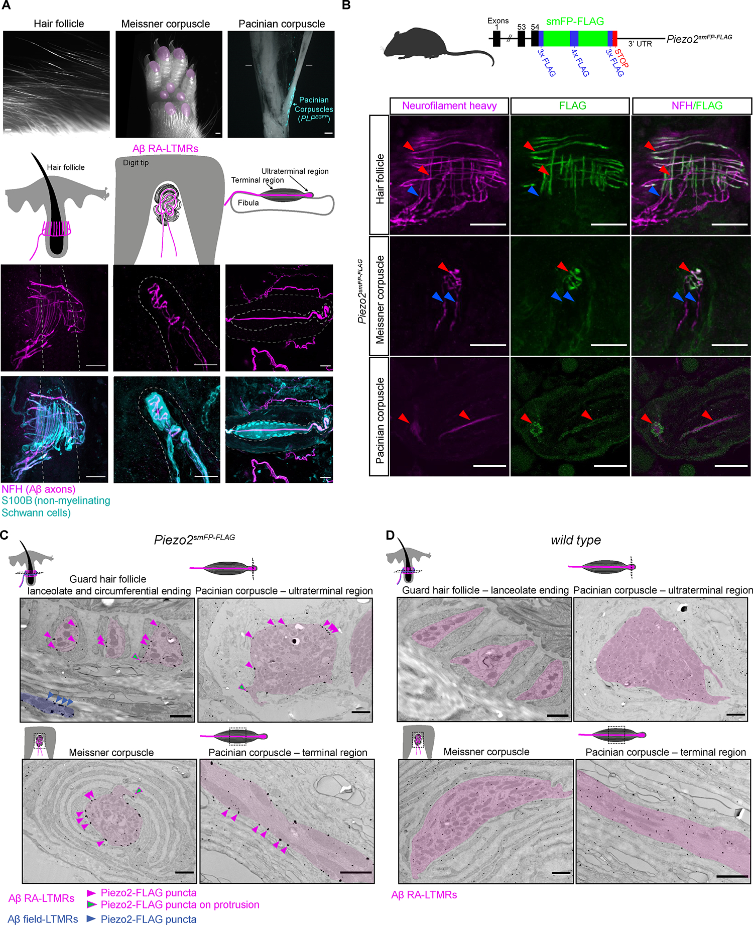
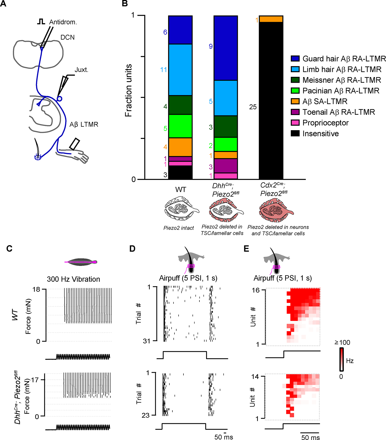
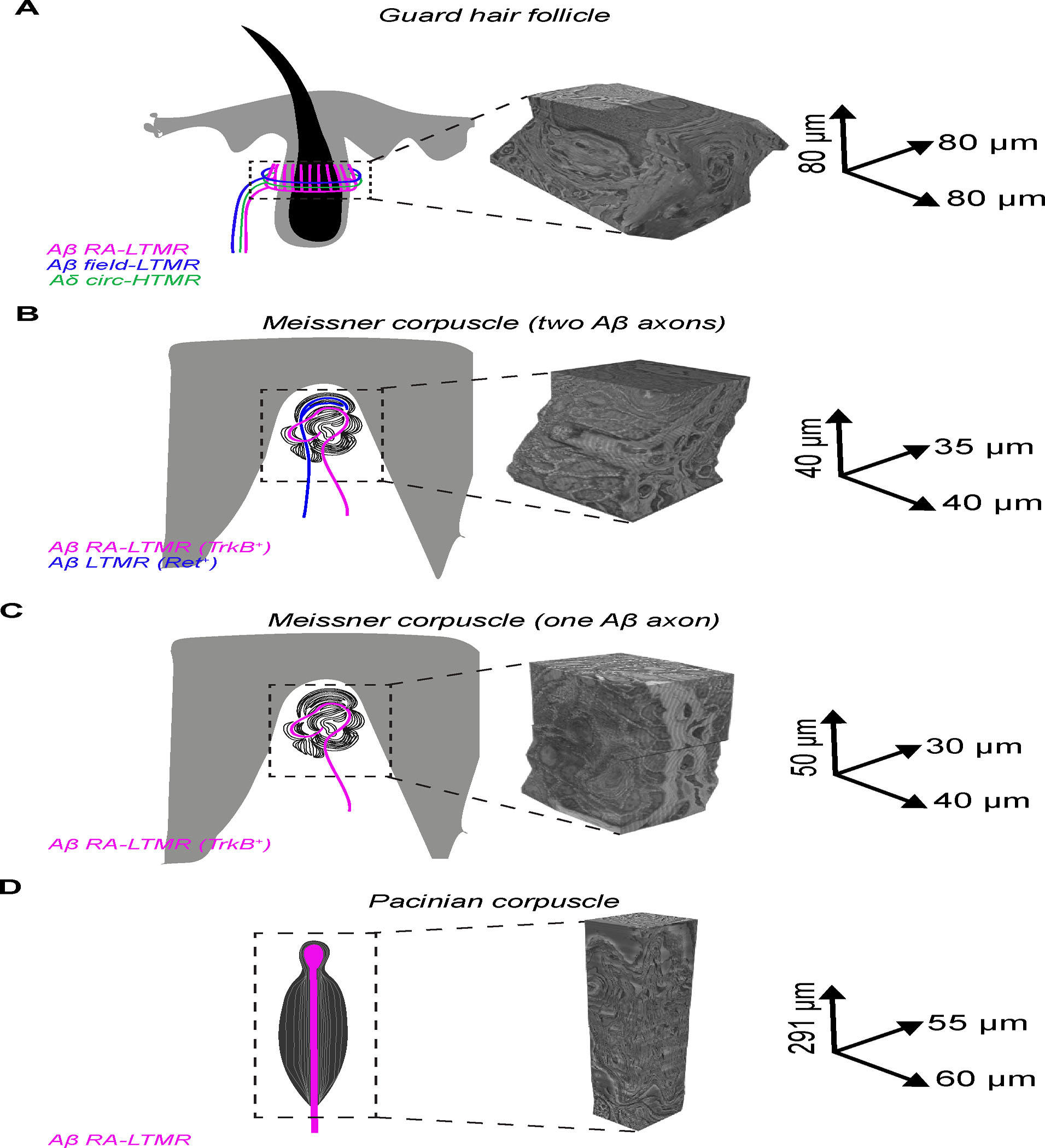
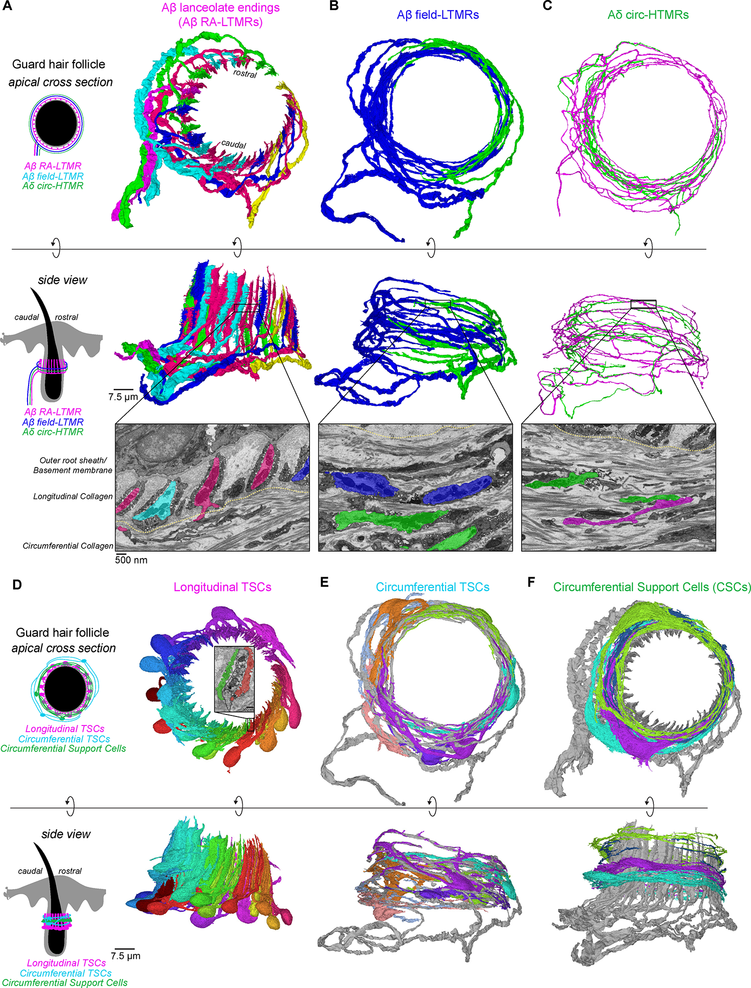
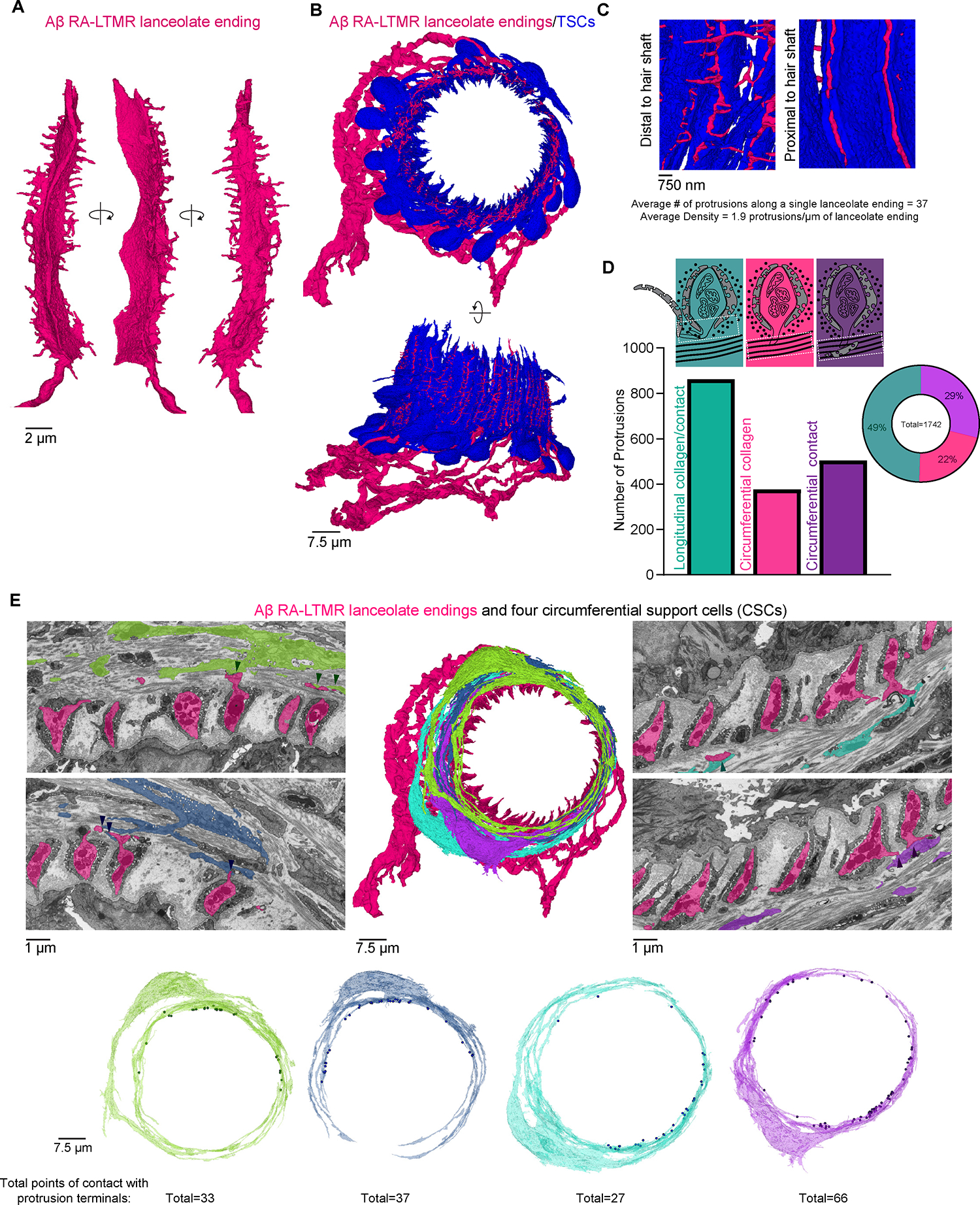
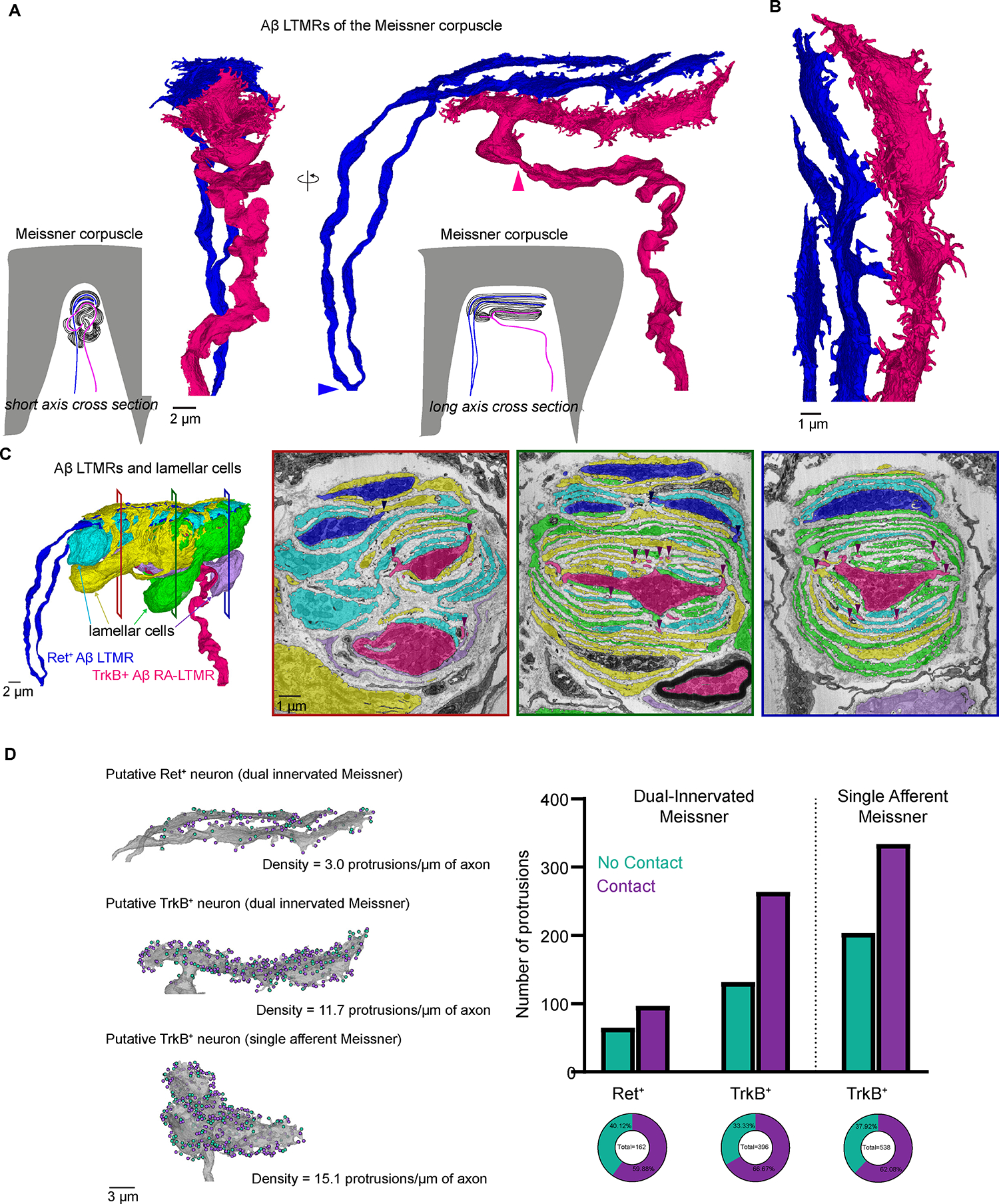
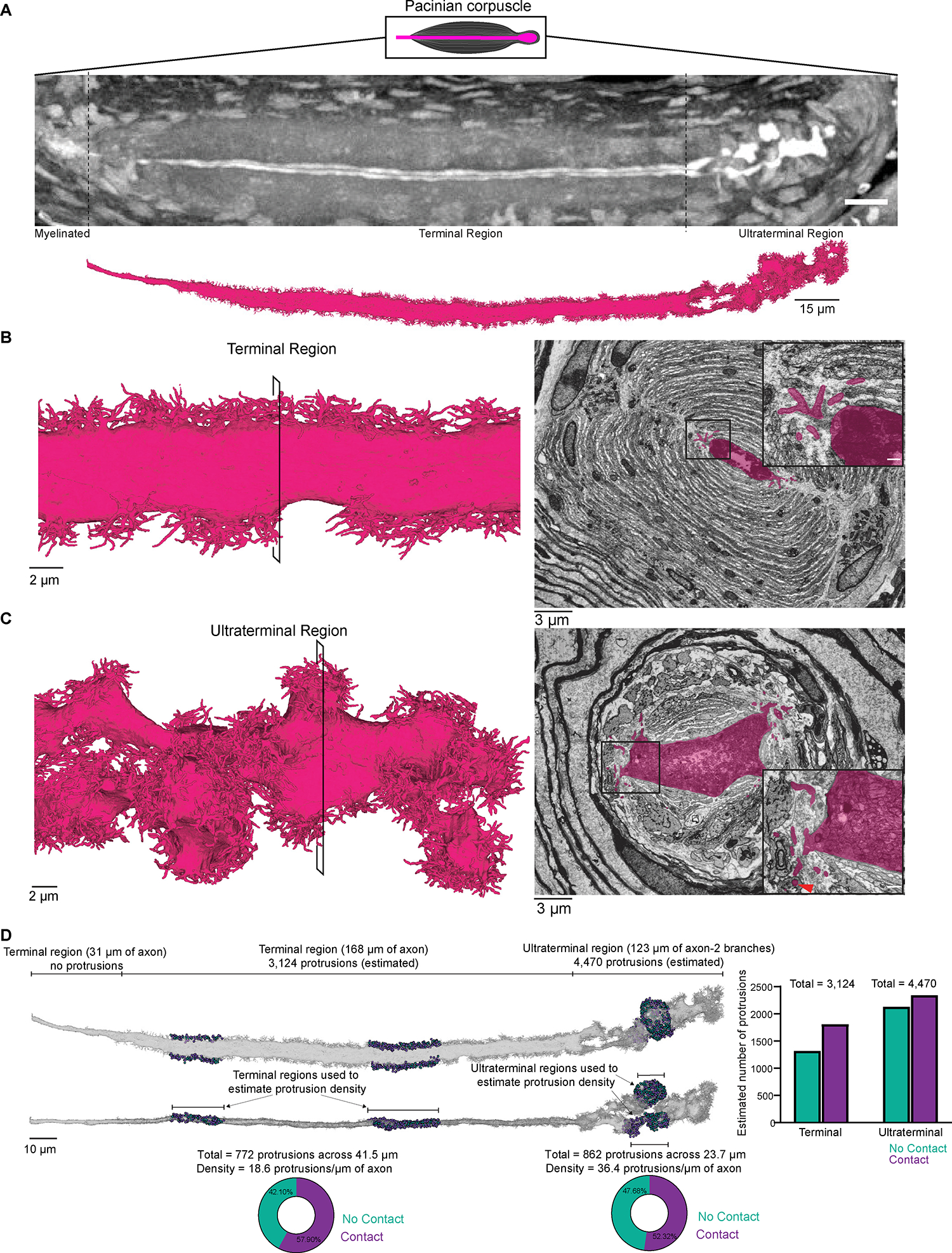
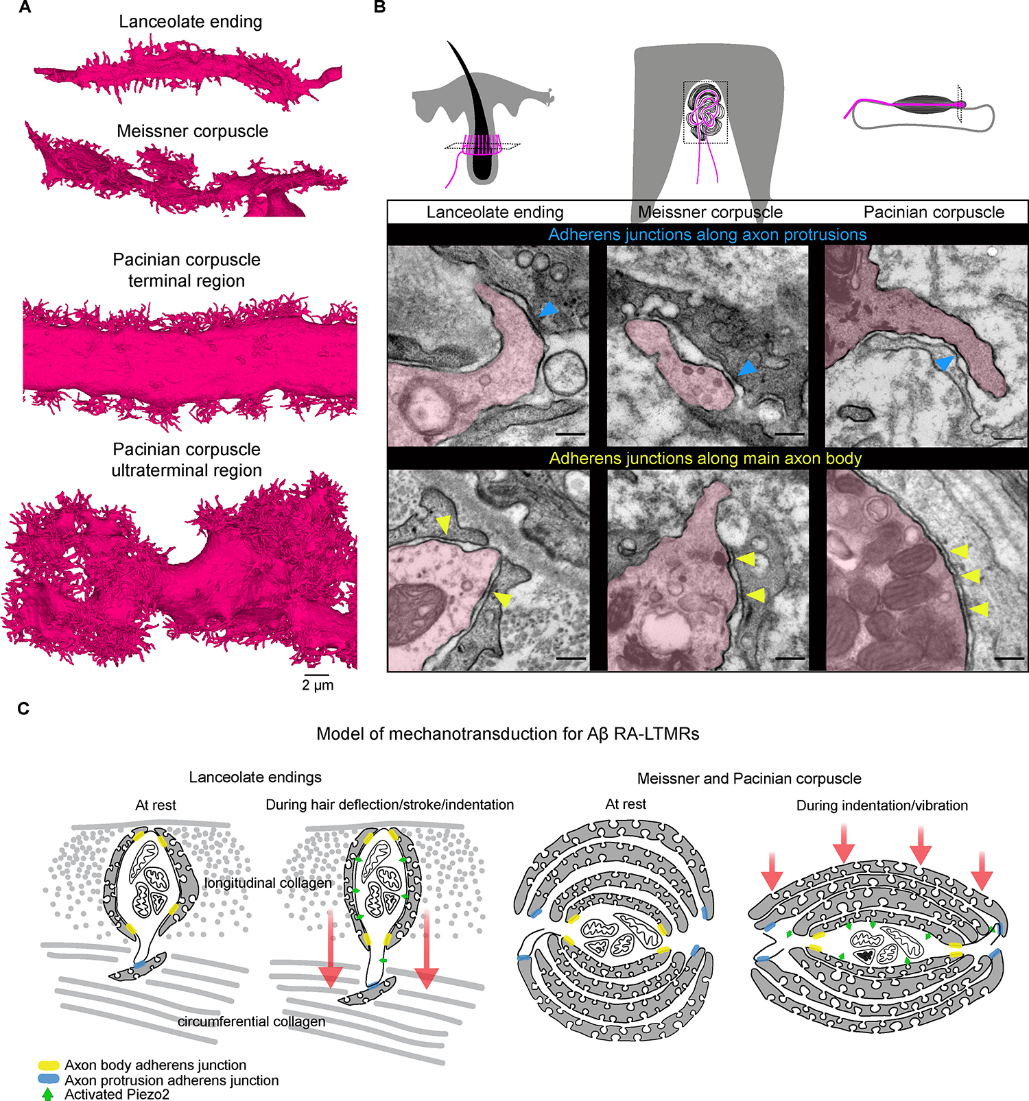
Across mammalian skin, structurally complex and diverse mechanosensory end organs respond to mechanical stimuli and enable our perception of dynamic, light touch. How forces act on morphologically dissimilar mechanosensory end organs of the skin to gate the requisite mechanotransduction channel Piezo2 and excite mechanosensory neurons is not understood. Here, we report high-resolution reconstructions of the hair follicle lanceolate complex, Meissner corpuscle, and Pacinian corpuscle and the subcellular distribution of Piezo2 within them. Across all three end organs, Piezo2 is restricted to the sensory axon membrane, including axon protrusions that extend from the axon body. These protrusions, which are numerous and elaborate extensively within the end organs, tether the axon to resident non-neuronal cells via adherens junctions. These findings support a unified model for dynamic touch in which mechanical stimuli stretch hundreds to thousands of axon protrusions across an end organ, opening proximal, axonal Piezo2 channels and exciting the neuron.

Keywords: Piezo2; focused ion beam scanning electron microscopy; light touch; mechanosensory neurons; mechanotransduction; touch end organs.


Conflict of interest statement

Declaration of interests C.S.X. is the inventor of a US patent assigned to HHMI for the enhanced FIB-SEM systems used in this work: Xu, C.S., Hayworth, K.J., and Hess, H.F. (2020) Enhanced FIB-SEM systems for large-volume 3D imaging. US Patent 10,600,615, 24 Mar. 2020.

Figures

Figure 1.
Figure 1.. Piezo2 is restricted to axonal endings across morphologically dissimilar Aβ RA-LTMRs.
(A) Image of three main skin/tissue regions that contain end organs formed by Aβ RA-LTMRs: lanceolate endings around hair follicles, Meissner corpuscles within glabrous skin of digit tips and pedal pads (regions indicated in pink), and Pacinian corpuscles (indicated by the PLPEGFP fluorescent label) around the periosteum of the fibula. Diagrams illustrate end organs with Aβ RA-LTMRs in magenta. Representative confocal images show Aβ RA-LTMRs (NFH+) and specialized non-neuronal Schwann cells (S100B+) of each end organ. Dashed lines outline the hair follicle (left), dermal papilla (middle), and boundary of outer and inner core (right). Scalebar, 20 μm. (B) Top, schematic diagram of the Piezo2smFP-FLAG allele. Bottom, confocal images of Piezo2-FLAG localization to Aβ RA-LTMR axons (NFH+) in the three end organs. Red arrowheads indicate co-localized FLAG and NFH signal. Blue arrowheads indicate axonal NFH signal outside the end organ that lacks FLAG signal. This experiment was repeated in two animals with littermate controls. Scalebar, 25 μm. (C) Immuno-electron micrographs from a Piezo2smFP-FLAG animal stained for FLAG with silver enhancement show localization of the Piezo2-FLAG fusion protein along the membranes of Aβ RA-LTMRs (pseudo-colored pink). Pink arrowheads indicate a subset of Piezo2-FLAG puncta along sensory axon membranes; green arrowheads indicate a subset of puncta along axon protrusions. Piezo2-FLAG is also localized to the membrane of Aβ field-LTMR circumferential endings around the guard hair (blue). This experiment was repeated in two animals with littermate controls. Scalebar, 1 μm. (D) Same as in (C) but for wild-type littermate of the Piezo2smFP-FLAG animal. See also Figures S1–S2.
Figure 2.
Figure 2.. Deletion of Piezo2 in TSCs and lamellar cells does not affect light touch responses in Aβ sensory neurons.
(A) Schematic of juxtacellular recordings. Aβ sensory neurons were identified with antidromic activation of the dorsal column nucleus (DCN) or dorsal column (DC). Mechanical stimuli were applied to the hindpaw and hairy thigh. (B) The fraction of DCN-projecting units identified as a specific sensory neuron class based on the location of receptive field and response to low-threshold mechanical stimuli in wild type (wt) animals (n=3), DhhCre;Piezo2fl/fl mice (n=3), and Cdx2Cre;Piezo2fl/fl mice (n=2). (C) Raster of single Pacinian Aβ RA-LTMR responses to vibration (300 Hz) at different forces in a wt (top) and a DhhCre;Piezo2fl/fl animal (bottom). (D) Same as in (C) but for hairy skin Aβ RA-LTMR responses to air puff. (E) Histograms of air puff responses for all Aβ hairy units activated by DCN stimulation in wt (top) and DhhCre;Piezo2fl/fl animals (bottom). In wt animals, 15/16 hairy units were air puff-sensitive. In the DhhCre;Piezo2fl/fl animals, 14/15 hairy units were air puff-sensitive. See also Figure S3.
Figure 3.
Figure 3.. Aligned FIB-SEM volumes of end organs formed by Aβ RA-LTMRs.
FIB-SEM volumes with global alignment correction and dimensions for skin samples containing (A) a single guard hair; (B) a Meissner corpuscle with two myelinated afferents; (C) a Meissner corpuscle with a single myelinated afferent; (D) a Pacinian corpuscle.
Figure 4.
Figure 4.. FIB-SEM reconstructions of Aβ and Aδ sensory neuron endings and associated non-neuronal cells of a guard hair follicle.
(A-C) 3D renderings of sensory neurons innervating a mouse guard hair follicle shown from an apical view (top panels) and from a side view (middle panels). Bottom panels: pseudo-colored images from FIB-SEM volume with the boundary between longitudinal and circumferential collagen matrix marked by the yellow line. (A) Six Aβ RA-LTMR axon segments form 47 lanceolate endings. (B) Two Aβ field-LTMRs and (C) two Aδ circ-HTMRs form circumferential endings. (D-F) 3D renderings of (D) 17 terminal Schwann cells (TSCs) within the longitudinal collagen that form intimate associations with lanceolate endings. Inset: an example of two different TSCs associated with the same lanceolate ending; (E) seven circumferential TSCs within the circumferential collagen matrix that associate with Aβ field-LTMRs (gray); (F) four circumferential support cells (CSCs) within the circumferential collagen matrix and frequently contact axon protrusions of Aβ RA-LTMR lanceolate endings (gray). See also Figure S4.
Figure 5.
Figure 5.. Axon protrusions along Aβ RA-LTMRs extend through TSC openings and contact CSCs within the guard hair’s circumferential collagen network.
(A) A single lanceolate ending with numerous axon protrusions shown from multiple perspectives. (B) Apical and side views of Aβ RA-LTMRs (magenta) and associated TSCs (blue). (C) Axon protrusions emerge at TSC gaps distal to the hair shaft. (D) Quantification of axon protrusion terminals. Teal: protrusions that terminate in local longitudinal collagen with or without making contact with local TSCs. Magenta: protrusions that extend into circumferential collagen but do not form cell contacts. Purple: protrusions that extend into circumferential collagen and form cell contacts. (E) Axon protrusions of Aβ RA-LTMRs that extend into the circumferential collagen network frequently contact four reconstructed CSCs (arrow heads). Points where each CSC contacts an axon protrusion are marked as dots on the 3D rendering of the cell. See also Figure S5.
Figure 6.
Figure 6.. FIB-SEM reconstructions of two Meissner corpuscles reveal the density of axon protrusions to be a structural correlate of Aβ LTMR tactile sensitivity.
(A) 3D renderings of two Aβ LTMRs that innervate a Meissner corpuscle. Arrowheads indicate the termination of myelination. (B) Higher magnification rendering of the terminal portion of the two Aβ LTMRs. (C) Left, 3D renderings of the two Aβ LTMRs and four lamellar cells that form the Meissner corpuscle. Right, pseudo-colored images from the FIB-SEM volume at different depths across the corpuscle. Arrowheads show points where axon protrusions contact lamellar cells. (D) Left, 3D renderings of the two axons from the dual-innervated corpuscle and of the axon from the single-innervated corpuscle. Teal dots indicate protrusions that terminate in the collagen (no contact). Purple dots indicate protrusions that contact lamellar cells. Right, quantification of protrusion terminals. See also Figure S6.
Figure 7.
Figure 7.. FIB-SEM reconstruction of a Pacinian corpuscle Aβ RA-LTMR reveals dense networks of axon protrusions that form contacts with lamellar cells.
(A) X-ray tomography of a Pacinian corpuscle (axon in white) prior to FIB-SEM imaging (top) and 3D rendering of the reconstructed Pacinian Aβ RA-LTMR after imaging (bottom). (B) Left, 3D rendering of a section of the Aβ RA-LTMR in the terminal region. Right, pseudo-colored image from the FIB-SEM volume with inset showing protrusions and their contacts with lamellar cells. (C) Same as in (B) except for the ultraterminal region. The red arrowhead indicates a protrusion encased by a lamellar cell at the inner and outer core boundary. (D) Characterization of protrusion terminals in the terminal and ultraterminal region. Two representative regions in the terminal and ultraterminal region (total of 4) were selected to quantify and characterize protrusion terminals. The pie charts show the percentage of protrusions within these regions that contact a lamellar cell (purple) or do not form a contact (teal). The statistics of protrusion terminals in these regions were used to estimate the total number of protrusions and their termination type within both the terminal and ultraterminal region (graphed on the right). See also Figure S7.
Figure 8.
Figure 8.. The conserved presence of adherens junctions along axon protrusion contacts with non-neuronal cells and a unified model of mechanotransduction.
(A) 3D renderings of a portion of the Aβ RA-LTMR axons from each end organ (terminal and ultraterminal region shown for the Pacinian corpuscle). Images are shown on the same scale. (B) TEM micrographs processed with tannic acid and post-stain showing adherens junctions between axon protrusions of Aβ RA-LTMRs (magenta) and resident non-neuronal cells (blue arrowheads) and along the main body of the Aβ RA-LTMRs and neighboring terminal Schwann cells or lamellar cells (yellow arrowheads) in each end organ. This experiment was repeated in two animals. Scalebar, 200 nm. (C) Schematic of proposed model of mechanotransduction for Aβ RA-LTMRs. Adherens junctions serve as anchor sites that render the Aβ RA-LTMR uniquely sensitive to dynamic stimuli. Hair deflection or skin indentation/vibration tugs on the hundreds to thousands of protrusions along the length of the axon within the end organ leading to stretching of the Aβ RA-LTMR membrane and activation of Piezo2. See also Figure S8.

Update of

Comment in

References

    1. Handler A, and Ginty DD (2021). The mechanosensory neurons of touch and their mechanisms of activation. Nat. Rev. Neurosci. 22, 521–537. 10.1038/s41583-021-00489-x. - DOI - PMC - PubMed
    1. Bai L, Lehnert BP, Liu J, Neubarth NL, Dickendesher TL, Nwe PH, Cassidy C, Woodbury CJ, and Ginty DD (2015). Genetic Identification of an Expansive Mechanoreceptor Sensitive to Skin Stroking. Cell 163, 1783–1795. 10.1016/j.cell.2015.11.060. - DOI - PMC - PubMed
    1. Neubarth NL, Emanuel AJ, Liu Y, Springel MW, Handler A, Zhang Q, Lehnert BP, Guo C, Orefice LL, Abdelaziz A, et al. (2020). Meissner corpuscles and their spatially intermingled afferents underlie gentle touch perception. Science (80-. ). 368, 1–12. 10.1126/science.abb2751. - DOI - PMC - PubMed
    1. Lewin GR, and McMahon SB (1991). Physiological properties of primary sensory neurons appropriately and inappropriately innervating skin in the adult rat. J. Neurophysiol. 66, 1205–1217. 10.1152/jn.1991.66.4.1205. - DOI - PubMed
    1. Koltzenburg M, Stucky CL, and Lewin GR (1997). Receptive properties of mouse sensory neurons innervating hairy skin. J. Neurophysiol. 78, 1841–1850. 10.1152/jn.1997.78.4.1841. - DOI - PubMed

Publication types

MeSH terms

LinkOut - more resources