. 2023 Dec;21(12):3477-3489.
doi: 10.1016/j.jtha.2023.09.004. Epub 2023 Sep 17.

Coagulation factor VIII regulates von Willebrand factor homeostasis invivo

Affiliations

Coagulation factor VIII regulates von Willebrand factor homeostasis invivo

Wenjing Cao et al. J Thromb Haemost. 2023 Dec.

Abstract

Background: Coagulation factor VIII (FVIII) and von Willebrand factor (VWF) circulate as a noncovalent complex, but each has its distinct functions. Binding of FVIII to VWF results in a prolongation of FVIII's half-life in circulation and modulates FVIII's immunogenicity during hemophilia therapy. However, the biological effect of FVIII and VWF interaction on VWF homeostasis is not fully understood.

Objectives: To determine the effect of FVIII in VWF proteolysis and homeostasis in vivo.

Methods: Mouse models, recombinant FVIII infusion, and patients with hemophilia A on a high dose FVIII for immune tolerance induction therapy or emicizumab for bleeding symptoms were included to address this question.

Results: An intravenous infusion of a recombinant B-domain less FVIII (BDD-FVIII) (40 and 160 μg/kg) into wild-type mice significantly reduced plasma VWF multimer sizes and its antigen levels; an infusion of a high but not low dose of BDD-FVIII into Adamts13+/- and Adamts13-/- mice also significantly reduced the size of VWF multimers. However, plasma levels of VWF antigen remained unchanged following administration of any dose BDD-FVIII into Adamts13-/- mice, suggesting partial ADAMTS-13 dependency in FVIII-augmented VWF degradation. Moreover, persistent expression of BDD-FVIII at ∼50 to 250 U/dL via AAV8 vector in hemophilia A mice also resulted in a significant reduction of plasma VWF multimer sizes and antigen levels. Finally, the sizes of plasma VWF multimers were significantly reduced in patients with hemophilia A who received a dose of recombinant or plasma-derived FVIII for immune tolerance induction therapy.

Conclusion: Our results demonstrate the pivotal role of FVIII as a cofactor regulating VWF proteolysis and homeostasis under various (patho)physiological conditions.

Keywords: ADAMTS-13; coagulation factor VIII; proteolysis; regulation; thrombosis; von Willebrand factor.


Conflict of interest statement

Declaration of competing interests X.L.Z. is a consultant and a member of the advisory boards for Alexion, Apollo, GC Biopharma, Sanofi, Stago, and Takeda. X.L.Z. is also the cofounder of Clotsolution. W.C. holds equity in Ivygen. All other authors have declared no relevant conflict.

Figures

Fig. 1.
Fig. 1.. Characterization of recombinant FVIII protein and mutant mice
. A. SDS-PAGE with Coomassie blue staining for a protein marker (Lane 1) and a purified recombinant B domain-deleted FVIII (DBBFVIII) (Lane 2); a, b, and c denote the intact, heavy chain, and light chain, respectively. B. Plasma ADAMTS13 activity in wild-type (WT), Adamts13+/− (A13+/−), and Adamts13−/− (A13−/−) mice determined by an ELISA-based assay using a pooled human normal plasma as a calibrator; C. Plasma FVIII activities in fVIII−/− mice 13 weeks following administration of nothing or AAV8-BDD-FVIII. Data in panels B and C were presented as the mean ± standard deviation (SD). Kruskal-Willis one way analysis and Mann-Whitney analysis were performed to determine the difference among three groups in panel B and between two groups in panel C. Here, four stars (****) indicates a p value <0.001.
Fig. 2.
Fig. 2.. Effect of recombinant BBD-FVIII on plasma VWF multimer distribution and antigen levels in wild type mice
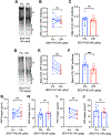
. A and B/C show the multimer distribution and the ratios of high (H) to low (L) molecular weight of VWF multimers, respectively, in wild-type (WT) mice prior to (pre) and 24 hours following an infusion of recombinant BDD-FVIII (40 μg/kg or 3.0 nM). D and E/F are the multimer distribution and the H/L ratios of VWF multimers, respectively, in WT mice pre and 24 hours of post infusion of BDD-FVIII (160 μg/kg or 12.1 nM). B/E. Paired individual values of H/L ratios of VWF multimers pre and post BDD-FVIII infusion. C/F. Individual values and mean ± SEM of H/L ratios of VWF multimers pre and post BDD-FVIII infusion. G/H and I/J show the plasma VWF antigen levels pre and 24 hours of post BDD-FVIII infusion at 40 μg/kg (3.0 nM) and 160 μg/kg (12.1 nM), respectively. G/I. Paired individual values of VWF antigen pre and post BDD-FVIII infusion. H/J. Individual values and mean ± SEM of VWF antigen pre and post BDD-FVIII infusion. A paired t-test was performed for determine the statistical significance of the difference of two different time points. Here *, **, and *** indicate a p value less than 0.05, 0.01, and 0.005, respectively.
Fig. 3.
Fig. 3.. Effect of recombinant BDD-FVIII on plasma VWF multimer distribution and its antigen levels in Adamts13+/− mice.
A and B/C show the multimer distribution and the ratios of high (H) to low (L) molecular weight of VWF multimers, respectively, in Adamts13+/− (A13+/−) mice pre and 24 hours post Infusion of BDD-FVIII at 40 μg/kg (or 3.0 nM). D and E/F are the multimer distribution and the ratios of H/L molecular weight of VWF multimers, respectively, in Adamts13+/− (A13+/−) mice pre and 24 hours post infusion of BDD-FVIII at 160 μg/kg (or 12.1 nM). G/H and I/J are the plasma VWF antigen levels pre and 24 hours of post infusion of BDD-FVIII at 40 μg/kg (or 3.0 nM) and 160 μg/kg (or 12.1 nM), respectively. A paired t-test was performed for determine the statistical significance of the difference of two different time points. Here, ns and **** indicate p value greater than 0.05 and less than 0.0001, respectively.
Fig. 4.
Fig. 4.. Effect of recombinant BDD-FVIII on plasma VWF multimer distribution and antigen in Adamts13−/− mice.
A and B/C show the plasma multimer distribution and the ratios of high (H) to low (L) molecular weight of VWF multimers, respectively, in Adamts13−/− (A13−/−) mice pre and 24 hours of post infusion of BDD-FVIII at 40 μg/kg (or 3.0 nM). D and E/F are the plasma multimer distribution and the ratios of H/L molecular weight of VWF multimers, respectively, in Adamts13−/− (A13−/−) mice pre and 24 hours post infusion of BDD-FVIII at 160 μg/kg (or 12.1 nM). G/H and I/J show the plasma VWF antigen levels in A13−/− mice pre and 24 hours of post infusion of BDD-FVIII at 40 μg/kg (or 3.0 nM) and 160 μg/kg (or 12.1 nM), respectively. A paired t-test was performed for determine the statistical significance between two different time points. Here, ns and *** indicate p value greater than 0.05 and less than 0.005, respectively.
Fig. 5.
Fig. 5.. Plasma VWF multimer distribution and antigen levels in fVIII−/− mice following AAV8-mediated expression of recombinant BDD-FVIII.
A. An outlined protocol for administration of AAV8-BDD-FVIII vector and blood collection. B and C. Plasma VWF multimer distribution in the untreated fVIII−/− mice (control) and mice received AAV8-BDD-FVIII (1×10¹¹ vg/mouse), respectively. D. The individual values and mean ± SEM of the ratios of high (H) to low (L) molecular weight of VWF multimers in fVIII−/− mice without or with expression of BDD-FVIII. E. The individual and mean ± SEM of plasma VWF antigen levels. Mann-Whitney test was performed to determine the statistical significance. Here, * and ** indicate the p value less than 0.05 and 0.01, respectively.
Fig. 6.
Fig. 6.. Plasma VWF multimer distribution and its antigen levels in haemophilia A patients with immune tolerance induction or emicizumab therapy.
A. A representative image showing plasma VWF multimer distribution in 4 haemophilia A patients (Pt1-Pt4) who were treated with (+) immune tolerance induction (ITI) or emicizumab (Emic.) or without (−). Plasma from 4 other haemophilia A patients without recent FVIII exposure (HA-82, HA-84, HA-85, and HA-86) was used for additional controls. B, C and D show the ratios of high (H) to low (L) molecular weights of plasma VWF multimers, VWF antigen levels, and FVIII antigen, respectively, in haemophilia A patients who did not have recent FVIII exposure (HA), those who treated with ITI, and those with Emici. The data are presented as the median and interquartile range (IQR). Kruskal-Wallis test was performed to determine the statistical significance. Here, ns, *, and ** indicate the p value greater than 0.05, less than 0.05, and 0.01, respectively.

References

    1. Wagner DD, Bonfanti R. von Willebrand factor and the endothelium. Mayo Clin Proc. 1991;66:621–627. - PubMed
    1. Mayadas TN, Wagner DD. von Willebrand factor biosynthesis and processing. Ann N Y Acad Sci. 1991;614:153–166. - PubMed
    1. Sadler JE. Biochemistry and genetics of von Willebrand factor. Annu Rev Biochem. 1998;67:395–424. - PubMed
    1. Dong JF, Moake JL, Bernardo A, et al. ADAMTS-13 metalloprotease interacts with the endothelial cell-derived ultra-large von Willebrand factor. J Biol Chem. 2003;278:29633–29639. - PubMed
    1. Dong JF, Moake JL, Nolasco L, et al. ADAMTS-13 rapidly cleaves newly secreted ultralarge von Willebrand factor multimers on the endothelial surface under flowing conditions. Blood. 2002;100:4033–4039. - PubMed

Publication types

LinkOut - more resources