. 2023 Sep 19;14(9):614.
doi: 10.1038/s41419-023-06131-1.

CSE reduces OTUD4 triggering lung epithelial cell apoptosis via PAI-1 degradation

Affiliations

CSE reduces OTUD4 triggering lung epithelial cell apoptosis via PAI-1 degradation

Lijuan Luo et al. Cell Death Dis. .

Abstract

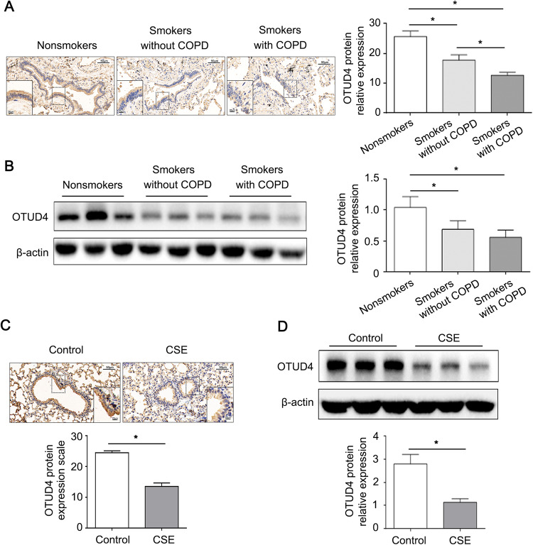
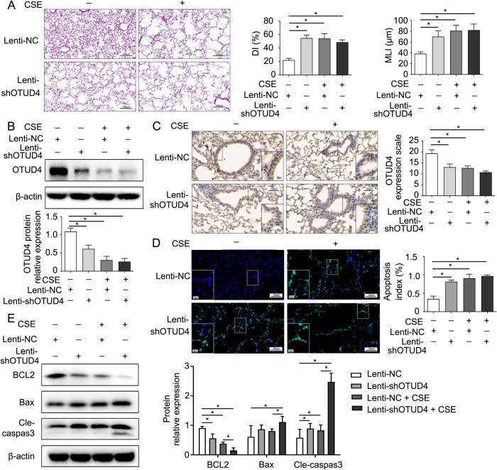
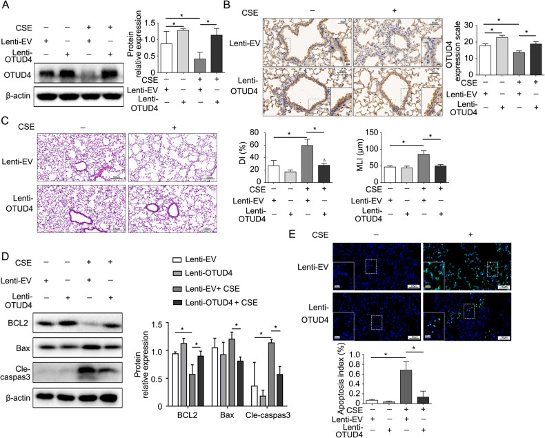
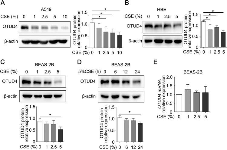
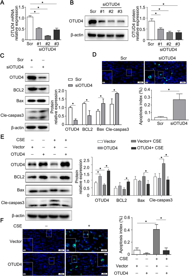
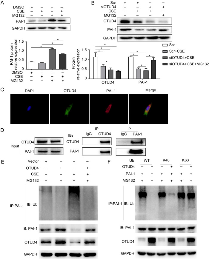
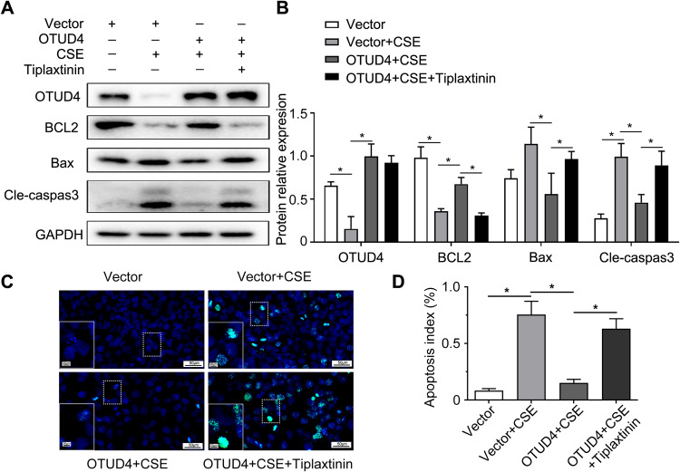
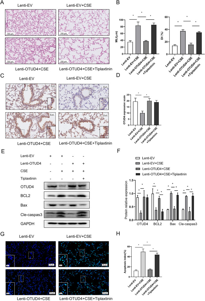
Ovarian tumor family deubiquitinase 4 (OTUD4), a member of the OTU deubiquitinating enzyme, is implicated to decrease in cancer to regulate cell apoptosis. However, the role of OTUD4 in cigarette smoke induced epithelial cell apoptosis and its mechanism have not been elucidated. In this study, we showed that OTUD4 protein reduced in CSE treated mice and airway epithelial cells. OTUD4 silence aggravated cell apoptosis and emphysematous change in the lung tissue of cigarette smoke extract (CSE) treated mice. Additionally, restoration of OTUD4 in the lung of mice alleviated CSE induced apoptosis and emphysematous morphology change. The effect of OTUD4 on cell apoptosis was also confirmed in vitro. Through protein profile screening, we identified that OTUD4 may interact with plasminogen activator inhibitor 1(PAI-1). We further confirmed that OTUD4 interacted with PAI-1 for de-ubiquitination and inhibiting CSE induced PAI-1 degradation. Furthermore, the protective role of OTUD4 in airway epithelial cells apoptosis was blocked by PAI-1 deactivation. Taken together, our data suggest that OTUD4 regulates cigarette smoke (CS)-triggered airway epithelial cell apoptosis via modulating PAI-1 degradation. Targeting OUTD4/PAI-1 signaling might potentially provide a therapeutic target against the lung cell apoptosis in cigarette smoke (CS)-induced emphysema.


Conflict of interest statement

The authors declare no competing interests.

Figures

Fig. 1
Fig. 1. OTUD4 is downregulated in the lung tissue of COPD patients and CSE-induced emphysema mouse model.
A Representative result of IHC (400× magnification) for OTUD4 in human lung tissue. B Western blotting to detect OTUD4 protein expression in human lung tissue. C OTUD4 was detected with IHC in the lung tissue of mice model. D Western blotting was applied to detect the OTUD4 protein expression in the lung tissue of mice. Data represents as mean ± SD of three independent experiments. *P < 0.05.
Fig. 2
Fig. 2. OTUD4 deficiency accelerates CSE induced emphysema via increasing airway epithelium apoptosis.
A Representative HE staining images (200×) of lung tissue were from control mice and OTUD4 deficiency mice. DI (%) and MLI (μm) were measured. Immunoblotting (B) and IHC (C) were used to detect OTUD4 protein expression (400×). D Apoptosis of lung cells in indicated group of mice were detected via TUNEL assay (400×). Apoptosis index (apoptotic cells/ total cells, %) was calculated. E BCL2, Bax and cleaved-caspase 3 protein expression was detected via immunoblotting. Lenti-NC: negative control; Lenti-shOTUD4: OTUD4 knockdown. Data were shown as mean ± SD of three independent experiments. *P < 0.05.
Fig. 3
Fig. 3. Enhanced OTUD4 alleviates emphysema by reducing cell apoptosis.
A Western blot to detect the expression of OTUD4 protein in the lung tissue of mice. B Representative results of IHC (400×) for OTUD4 in different groups were shown. C Histological changes of lung sections were shown with H&E (200×) staining. Morphometric measurements of MLI (μm) and DI (%) were plotted. D The expression of apoptotic protein including BCL2, Bax and cleaved caspase 3 were detected in the lung tissue of mice. E Apoptotic nuclei were detected by TUNEL staining (400×) in the lung tissue of mice. Lenti-EV: empty vector, Lenti-OTUD4: OTUD4 overexpression; Data were shown as mean ± SD of three independent experiments. *P < 0.05.
Fig. 4
Fig. 4. OTUD4 decreases in CSE induced lung epithelial cells.
AD OTUD4 relative expression was assayed by western blotting in type II epithelial A549 cells, HBEs and BEAS-2b. E RT-qPCR to detect the mRNA level of OTUD4 in CSE treated BEAS-2b cells. Data represents as mean ± SD of three independent experiments. *P < 0.05.
Fig. 5
Fig. 5. OTUD4 regulates apoptosis in lung epithelial cells induced by CSE.
A, B The mRNA and protein level of OTUD4 were confirmed with RT-qPCR and western blot. C OTUD4, BCL2, Bax, cleaved-caspase 3 or β-actin were assayed with western blotting after OTUD4 knockdown. D TUNEL staining (400×) to detect apoptotic nuclei of BEAS-2b cells. E Western blot to detect the protein level of BCL2, Bax and cleaved caspase 3. F Apoptotic nuclei of BEAS-2b cells were detected by TUNEL staining (400×). Data were shown as mean ± SD of three independent experiments. *p < 0.05.
Fig. 6
Fig. 6. OTUD4 deubiquitinates PAI-1 and inhibits its proteasomal degradation.
A Western-blot assayed the protein level of PAI-1 in BEAS-2b cells. B PAI-1 protein expression in the lung tissue of mice was detected by western blotting. C CHX or MG132 was subjected to BEAS-2b cells. PAI-1 is with a half-life of about 3.3 h. D, E PAI-1 protein expression was detected by western blotting in BEAS-2b cells after OTUD4 knockdown or overexpression with CHX treatment. Data represents as mean ± SD of three independent experiments. *P < 0.05.
Fig. 7
Fig. 7. OTUD4 deubiquitinates PAI-1 and inhibits its proteasomal degradation.
A, B BEAS-2b cells were treated with CSE or MG132, CSE or MG132 was subjected to BEAS-2b cells after OTUD4 knockdown, Western blotting was used to detect OTUD4 and PAI-1 protein level. C Immunofluorescence stanning (400×) of OTUD4 (green), PAI-1 (red) and DAPI (blue) and merged image (upper panel). D Immunoprecipitation of OTUD4 and PAI-1 (lower panel). E Ubiquitination assay of PAI-1 in CSE induced BEAS-2b cells after OTUD4 overexpression. F WT, K48, or K63 Ub was co-transfected with PAI-1 and OTUD4 into HEK293T cells. After treatment with 5 μM MG132 for 5 h, cell lysates were subjected to ubiquitination assay. Data represents as mean ± SD of three independent experiments. *P < 0.05.
Fig. 8
Fig. 8. Inhibition of PAI-1 perturbs the protective effect of OTUD4 on CSE induced apoptosis in BEAS-2b cells.
A OTUD4 overexpressed cells were treated with PAI-1 inhibitor (tiplaxitinin) or CSE. OTUD4, BCL2, Bax, and Cleaved-caspase3 were detected by western blot. B Densitometry of OTUD4, BCL2, Bax, Cleaved-caspase3. C Apoptotic nuclei were detected by TUNEL staining (400×) in OTUD4 overexpressed BEAS-2b cells treated with CSE or tiplaxitinin. D Apoptotic index (%) was plotted. Data represents as mean ± SD of three independent experiments. *P < 0.05.
Fig. 9
Fig. 9. Inhibition of PAI-1 perturbs the protective effect of OTUD4 on CSE induced apoptosis in lung tissue of mice.
A, B Histological changes of lung sections were shown with H&E (200×) staining. Morphometric measurements of MLI (μm) and DI (%) were plotted. C-D Representative results of IHC (400×) for OTUD4 in different groups were shown. E, F Western blot to detect the expression of OTUD4, BCL2, Bax and cleaved caspase 3 protein in the lung tissue of mice. G, H Apoptotic nuclei were detected by TUNEL staining (400×) in the lung tissue of mice. Lenti-EV: empty vector, Lenti-OTUD4: OTUD4 overexpression; Data were shown as mean ± SD of three independent experiments. *P < 0.05.

Similar articles

Cited by

References

    1. Halpin DMG, Celli BR, Criner GJ, Frith P, López Varela MV, Salvi S, et al. The GOLD Summit on chronic obstructive pulmonary disease in low- and middle-income countries. Int J Tuberc Lung Dis. 2019;23:1131–41.. doi: 10.5588/ijtld.19.0397. - DOI - PubMed
    1. Heron M. Deaths: leading causes for 2008. Natl Vital– Stat Rep. : Cent Dis Control Prev, Natl Cent Health Stat, Natl Vital– Stat Syst. 2012;60:1–94. - PubMed
    1. Barnes PJ, Shapiro SD, Pauwels RA. Chronic obstructive pulmonary disease: molecular and cellular mechanisms. Eur Respir J. 2003;22:672–88. doi: 10.1183/09031936.03.00040703. - DOI - PubMed
    1. Slebos DJ, Ryter SW, van der Toorn M, Liu F, Guo F, Baty CJ, et al. Mitochondrial localization and function of heme oxygenase-1 in cigarette smoke-induced cell death. Am J Respir Cell Mol Biol. 2007;36:409–17. doi: 10.1165/rcmb.2006-0214OC. - DOI - PMC - PubMed
    1. Yoshida M, Minagawa S, Araya J, Sakamoto T, Hara H, Tsubouchi K, et al. Involvement of cigarette smoke-induced epithelial cell ferroptosis in COPD pathogenesis. Nat Commun. 2019;10:3145. doi: 10.1038/s41467-019-10991-7. - DOI - PMC - PubMed

Publication types

Substances