. 2023 Sep 19;6(1):956.
doi: 10.1038/s42003-023-05334-8.

Deep learning driven de novo drug design based on gastric proton pump structures

Affiliations

Deep learning driven de novo drug design based on gastric proton pump structures

Kazuhiro Abe et al. Commun Biol. .

Abstract

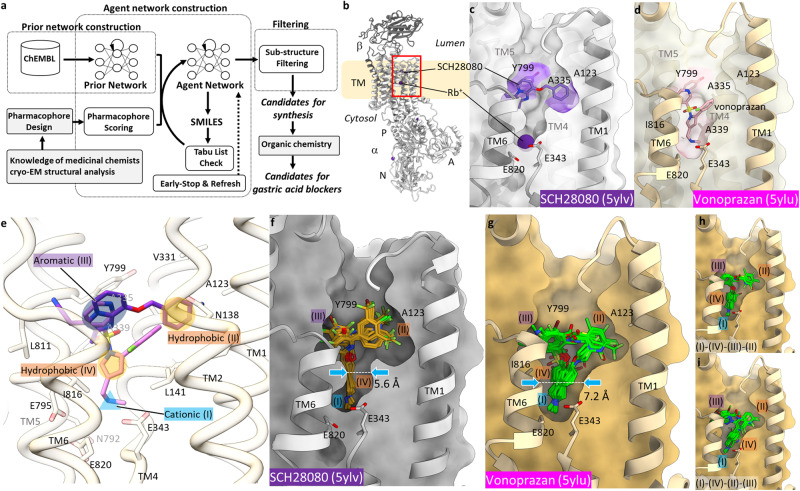
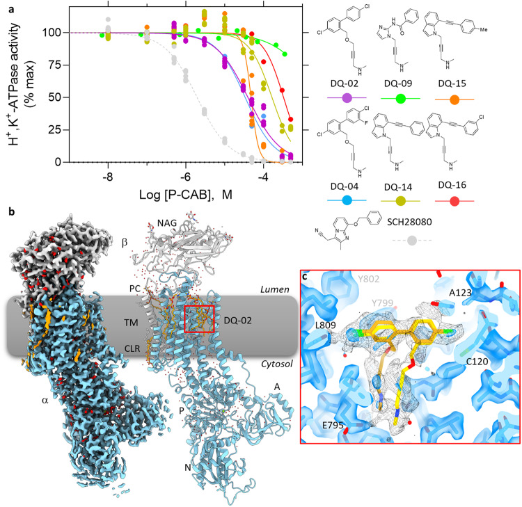
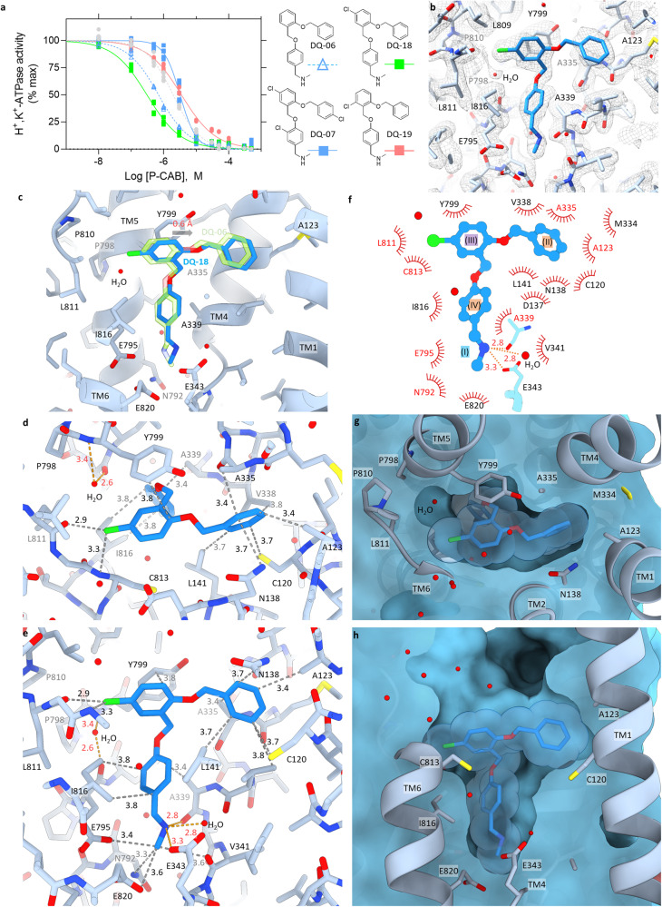
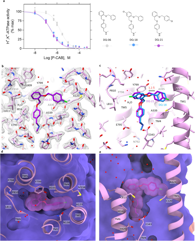
Existing drugs often suffer in their effectiveness due to detrimental side effects, low binding affinity or pharmacokinetic problems. This may be overcome by the development of distinct compounds. Here, we exploit the rich structural basis of drug-bound gastric proton pump to develop compounds with strong inhibitory potency, employing a combinatorial approach utilizing deep generative models for de novo drug design with organic synthesis and cryo-EM structural analysis. Candidate compounds that satisfy pharmacophores defined in the drug-bound proton pump structures, were designed in silico utilizing our deep generative models, a workflow termed Deep Quartet. Several candidates were synthesized and screened according to their inhibition potencies in vitro, and their binding poses were in turn identified by cryo-EM. Structures reaching up to 2.10 Å resolution allowed us to evaluate and re-design compound structures, heralding the most potent compound in this study, DQ-18 (N-methyl-4-((2-(benzyloxy)-5-chlorobenzyl)oxy)benzylamine), which shows a Ki value of 47.6 nM. Further high-resolution cryo-EM analysis at 2.08 Å resolution unambiguously determined the DQ-18 binding pose. Our integrated approach offers a framework for structure-based de novo drug development based on the desired pharmacophores within the protein structure.


Conflict of interest statement

A.Y. is a CEO of Institute for Theoretical Medicine, Inc. C.K. is an employee of INTAGE Healthcare Inc. Other authors declare no competing interests.

Figures

Fig. 1
Fig. 1. De novo drug design based on the desired pharmacophores.
a Workflow of “Deep Quartet”. See main text and “Methods” for details. b Crystal structure of the gastric H+,K+-ATPase complexed with SCH28080 (PDB ID: 5YLV, gray) in ribbon representation, viewed parallel to the membrane plane with its luminal-side facing upwards. c, d Close-up view of the P-CAB-binding site (indicated as a red box in b). Clipped cross section of the luminal-facing conduit (surface) of SCH28080- (gray) and vonoprazan- (PDB ID: 5YLU, wheat) bound forms from the viewpoint similar to (b). Positions of P-CAB binding are indicated as purple or pink surface for SCH28080 or vonoprazan, respectively. e Defined pharmacophore features (I–IV, see text for details) on the luminal-facing cavity of the H+,K+-ATPase structure. Navy and yellow ovals, and blue triangle indicates pharmacophore features with aromatic, hydrophobic or cationic properties, respectively. f Binding poses of candidate compounds in the template structure (5YLV) calculated by Deep Quartet. g Same as (f) but with vonoprazan-bound form used as a template (5YLU). The most constrained positions of the lumina-facing conduit in each structure are indicated as dotted lines and blue arrows, with accompanying distance in Å. Two distinct binding modes of candidates from vonoprazan-bound form are displayed separately in (h) and (i). In (c, d, fi), TM2 is omitted for clarity.
Fig. 2
Fig. 2. Inhibition potency and the binding pose of DQ-02.
a Dose-dependent inhibition of H+,K+-ATPase activity by indicated synthesized compounds (SCH28080 as a control, gray circles and a dashed line). Data plotted represent each data point from triplicate of three independent measurement at 12 different concentrations of P-CABs, using membrane fractions purified from pig stomach. b EM potential map (colored surface) and cartoon model of the gastric H+,K+-ATPase complexed with DQ-02 (α-subunit, skyblue; β-subunit, gray; lipids, orange; waters, red). c Close-up view of the DQ-02 binding site indicated by the red box in (b). Transparent blue surface and black mesh represent EM potential maps in high and low-contour levels, respectively. Only the EM density around DQ-02 is displayed for low-contour map (mesh). Two possible conformations of DQ-02 (gold and yellow sticks) are shown.
Fig. 3
Fig. 3. Inhibition potency and the binding pose of DQ-06.
a Dose-dependent inhibition of H+,K+-ATPase activity by indicated compounds as shown in Fig. 2a. Data plotted represent each data point from triplicate of three independent measurement at 12 different concentrations of P-CABs. Three benzene rings in DQ-06 are attributed as illustrated in the figure, which corresponds to the defined pharmacophore features (II)–(IV). b EM potential map (mesh) and the atomic model of the gastric H+,K+-ATPase complexed with DQ-06 (green sticks). Shown is a close-up view of the DQ-06 binding site as in Fig. 2c. c, d Molecular interactions between H+,K+-ATPase and DQ-06 in stick representation. Hydrophilic and hydrophobic atoms of DQ-06 within 3.5 and 4.0 Å distance from amino acids of H+,K+-ATPase are connected by orange and gray dotted lines, respectively. Panels are viewed from luminal side (c), or parallel to the membrane plane with luminal-side up (d). e, f Clipped cross sections of the DQ-06 binding site from the viewpoints approximately similar to (c) and (d). Molecular surface (gray) of H+,K+-ATPase shows the dimension of the binding site in which DQ-06 is accommodated (green stick with transparent van der Waals spheres). Except for TM2, which is removed from the figure for clarity, TM helices and some of the key amino acids are shown in ribbon and stick representations. g A schematic 2D representation of DQ-06 binding pose. Hydrophobic residues that are located within 3.9 Å from DQ-06 are shown, and those within 3.5 Å are highlighted as red. Expected polar interactions within 3.5 Å are indicated as orange dotted lines.
Fig. 4
Fig. 4. Inhibition potency and the binding pose of DQ-18.
a Dose-dependent inhibition of H+,K+-ATPase activity by indicated P-CABs (DQ-06, 07, 18, 19 and SCH28080) as shown in Fig. 2a. Data plotted represent each data point from triplicate of three independent measurement at 12 different concentrations of P-CABs. b EM potential map (mesh) and atomic model of the gastric H+,K+-ATPase complexed with DQ-18 (blue sticks). c Comparison of the binding poses between DQ-06 (green) and DQ-18 (blue). Arrow indicates the displacement of the binding position from DQ-06 to DQ-18 (0.6 Å). d, e Molecular interactions between H+,K+-ATPase and DQ-18 in stick representation as shown in Fig. 3c, d. f A schematic 2D representation of DQ-18 binding pose as shown in Fig. 3g. g, h Clipped cross sections of the DQ-18 binding site as in Fig. 3e, f.
Fig. 5
Fig. 5. Inhibition potency and the binding pose of DQ-21.
a Dose-dependent inhibition of H+,K+-ATPase activity by indicated P-CABs (DQ-06, 18 and 21) as shown in Fig. 2a. Data plotted represent each data point from triplicate of three independent measurement at 12 different concentrations of P-CABs. b EM potential map (mesh) and atomic model of the gastric H+,K+-ATPase complexed with DQ-21 (purple sticks). c Comparison of the binding poses between DQ-18 (blue) and DQ-21 (purple). Arrow indicates the displacement of the binding position from DQ-18 to DQ-21 (1.2 Å). d, e Clipped cross sections of the DQ-21 binding site as in Fig. 3e, f.

Similar articles

Cited by

References

    1. Tanaka S, et al. Structural basis for binding of potassium-competitive acid blockers to the gastric proton pump. J. Med. Chem. 2022;65:7843–7853. - PubMed
    1. Abe K, Irie K, Nakanishi H, Suzuki H, Fujiyoshi Y. Crystal structures of the gastric proton pump. Nature. 2018;556:214–229. - PubMed
    1. Shin JM, Sachs G. Pharmacology of proton pump inhibitors. Curr. Gastroenterol. Rep. 2008;10:528–534. - PMC - PubMed
    1. Wallace JL. How do NSAIDs cause ulcer disease? Best Pract. Res. Clin. Gastroenterol. 2000;14:147–159. - PubMed
    1. Hain E, Coriat R, Dousset B, Gaujoux S. Prise en charge du gastrinome. Presse Med. 2016;45:986–991. - PubMed

Publication types