

Impact of myocardial injury on regional left ventricular function in the course of acute myocarditis with preserved ejection fraction: insights from segmental feature tracking strain analysis using cine cardiac MRI
- PMID: 37726513
- PMCID: PMC9797452
- DOI: 10.1007/s10554-022-02601-3
Impact of myocardial injury on regional left ventricular function in the course of acute myocarditis with preserved ejection fraction: insights from segmental feature tracking strain analysis using cine cardiac MRI
Abstract
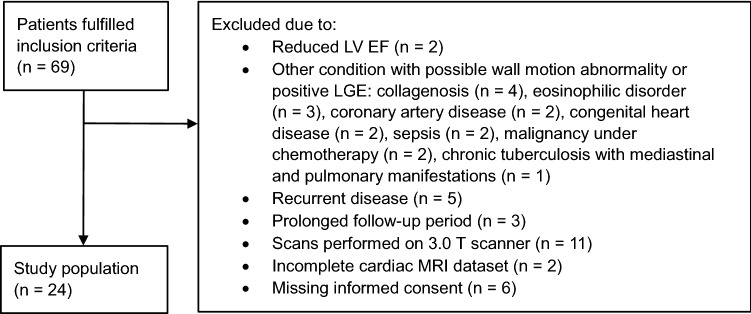
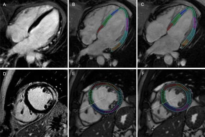
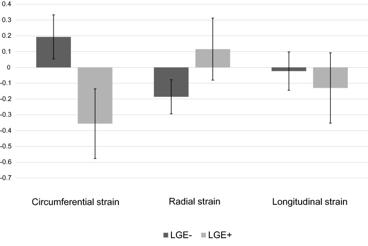
The aim of this study was to provide insights into myocardial adaptation over time in myocyte injury caused by acute myocarditis with preserved ejection fraction. The effect of myocardial injury, as defined by the presence of late gadolinium enhancement (LGE), on the change of left ventricular (LV) segmental strain parameters was evaluated in a longitudinal analysis. Patients with a first episode of acute myocarditis were enrolled retrospectively. Peak radial (PRS), longitudinal (PLS) and circumferential (PCS) LV segmental strain values at baseline and at follow-up were computed using feature tracking cine cardiac magnetic resonance imaging. The change of segmental strain values in LGE positive (LGE+) and LGE negative (LGE-) segments was compared over a course of 89 ± 20 days. In 24 patients, 100 LGE+ segments and 284 LGE- segments were analysed. Between LGE+ and LGE- segments, significant differences were found for the change of segmental PCS (p < 0.001) and segmental PRS (p = 0.006). LGE + segments showed an increase in contractility, indicating recovery, and LGE- segments showed a decrease in contractility, indicating normalisation after a hypercontractile state or impairment of an initially normal contracting segment. No significant difference between LGE+ and LGE- segments was found for the change in segmental PLS. In the course of acute myocarditis with preserved ejection fraction, regional myocardial function adapts inversely in segments with and without LGE. As these effects seem to counterbalance each other, global functional parameters might be of limited use in monitoring functional recovery of these patients.
Keywords: Cardiac magnetic resonance imaging; Feature tracking strain analysis; Late gadolinium enhancement; Myocardial strain; Myocarditis.
© 2022. The Author(s).
Conflict of interest statement
The authors declared no conflicts of interest.
Figures
Comment in
-
Cardiac magnetic resonance strain imaging in acute myocarditis with preserved ejection fraction: promising and evolving.Int J Cardiovasc Imaging. 2022 Aug;38(8):1863-1864. doi: 10.1007/s10554-022-02630-y. Epub 2022 Jun 21. Int J Cardiovasc Imaging. 2022. PMID: 37726520 No abstract available.
Similar articles
-
Cardiac magnetic resonance feature tracking myocardial strain analysis in suspected acute myocarditis: diagnostic value and association with severity of myocardial injury.BMC Cardiovasc Disord. 2023 Mar 28;23(1):162. doi: 10.1186/s12872-023-03201-2. BMC Cardiovasc Disord. 2023. PMID: 36977995 Free PMC article.
-
Diagnostic and Prognostic Value of Cardiac Magnetic Resonance Strain in Suspected Myocarditis With Preserved LV-EF: A Comparison Between Patients With Negative and Positive Late Gadolinium Enhancement Findings.J Magn Reson Imaging. 2022 Apr;55(4):1109-1119. doi: 10.1002/jmri.27873. Epub 2021 Aug 8. J Magn Reson Imaging. 2022. PMID: 34369030
-
Two-dimensional speckle-tracking-derived segmental peak systolic longitudinal strain identifies regional myocardial involvement in patients with myocarditis and normal global left ventricular systolic function.Pediatr Cardiol. 2015 Jun;36(5):950-9. doi: 10.1007/s00246-015-1105-9. Epub 2015 Jan 24. Pediatr Cardiol. 2015. PMID: 25617227
-
Impact of Myocardial Fibrosis on Left Ventricular Function Evaluated by Feature-Tracking Myocardial Strain Cardiac Magnetic Resonance in Competitive Male Triathletes With Normal Ejection Fraction.Circ J. 2019 Jun 25;83(7):1553-1562. doi: 10.1253/circj.CJ-18-1388. Epub 2019 May 11. Circ J. 2019. PMID: 31080228 Clinical Trial.
-
Comprehensive Cardiovascular Magnetic Resonance-Derived Myocardial Strain Analysis Provides Independent Prognostic Value in Acute Myocarditis.J Am Heart Assoc. 2022 Oct 4;11(19):e025106. doi: 10.1161/JAHA.121.025106. Epub 2022 Sep 21. J Am Heart Assoc. 2022. PMID: 36129042 Free PMC article.
Cited by
-
Effect of late gadolinium enhancement on left atrial impairment in myocarditis patients.Eur Radiol. 2024 Mar;34(3):1846-1853. doi: 10.1007/s00330-023-10176-3. Epub 2023 Sep 2. Eur Radiol. 2024. PMID: 37658889 Free PMC article.
-
Left ventricle strain and T1 mapping evaluation in a mouse model with myocardial infarction.Sci Rep. 2025 Aug 12;15(1):29579. doi: 10.1038/s41598-025-09699-0. Sci Rep. 2025. PMID: 40796920 Free PMC article.
-
Comparative analysis of late gadolinium enhancement assessment techniques for monitoring fibrotic changes in myocarditis follow-up.Eur Radiol. 2024 Nov;34(11):7264-7274. doi: 10.1007/s00330-024-10756-x. Epub 2024 May 4. Eur Radiol. 2024. PMID: 38703188 Free PMC article.
References
-
- Francone M, Chimenti C, Galea N, Scopelliti F, Verardo R, Galea R, Carbone I, Catalano C, et al. CMR sensitivity varies with clinical presentation and extent of cell necrosis in biopsy-proven acute myocarditis. JACC Cardiovasc Imaging. 2014;7(3):254–263. doi: 10.1016/j.jcmg.2013.10.011. - DOI - PubMed
-
- Jereczek-Fossa BA, Surgo A, Maisonneuve P, Maucieri A, Gerardi MA, Zerini D, Marvaso G, Ciardo D, et al. Late toxicity of image-guided hypofractionated radiotherapy for prostate: non-randomized comparison with conventional fractionation. Radiol Med. 2019;124(1):65–78. doi: 10.1007/s11547-018-0937-9. - DOI - PubMed
-
- Nucifora G, Miani D, Di Chiara A, Piccoli G, Artico J, Puppato M, Slavich G, De Biasio M, et al. Infarct-like acute myocarditis: relation between electrocardiographic findings and myocardial damage as assessed by cardiac magnetic resonance imaging. Clin Cardiol. 2013;36(3):146–152. doi: 10.1002/clc.22088. - DOI - PMC - PubMed
-
- Ammirati E, Moroni F, Sormani P, Peritore A, Milazzo A, Quattrocchi G, Cipriani M, Oliva F, et al. Quantitative changes in late gadolinium enhancement at cardiac magnetic resonance in the early phase of acute myocarditis. Int J Cardiol. 2017;231:216–221. doi: 10.1016/j.ijcard.2016.11.282. - DOI - PubMed
MeSH terms
Substances
LinkOut - more resources
Full Text Sources
Research Materials
