

Cryo EM structures map a post vaccination polyclonal antibody response to canine parvovirus
- PMID: 37726539
- PMCID: PMC10509169
- DOI: 10.1038/s42003-023-05319-7
Cryo EM structures map a post vaccination polyclonal antibody response to canine parvovirus
Abstract
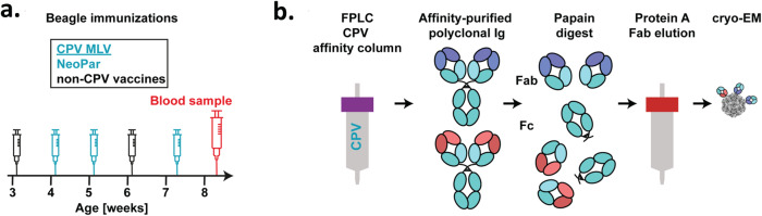
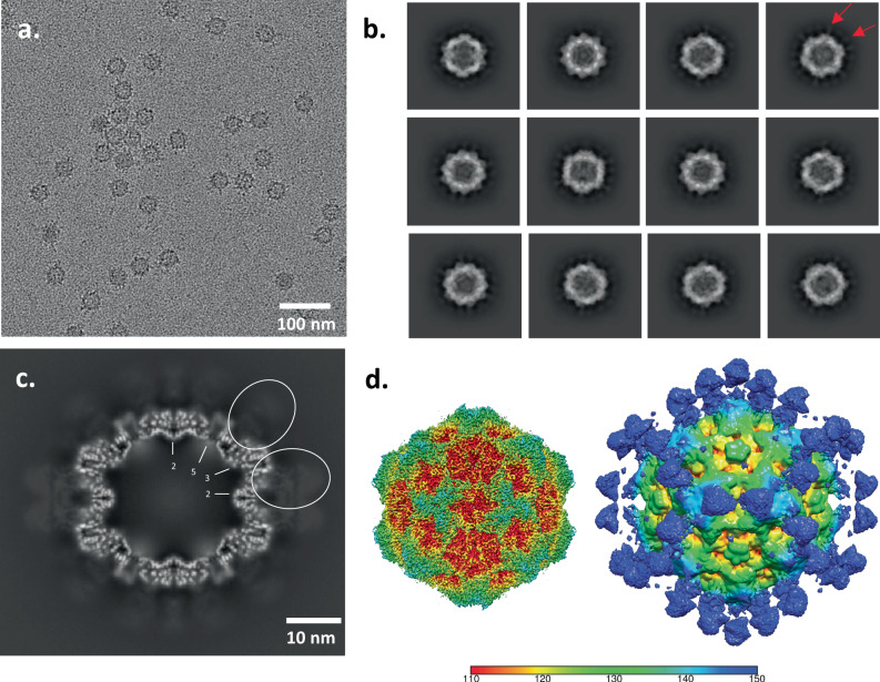
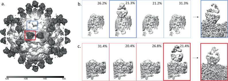
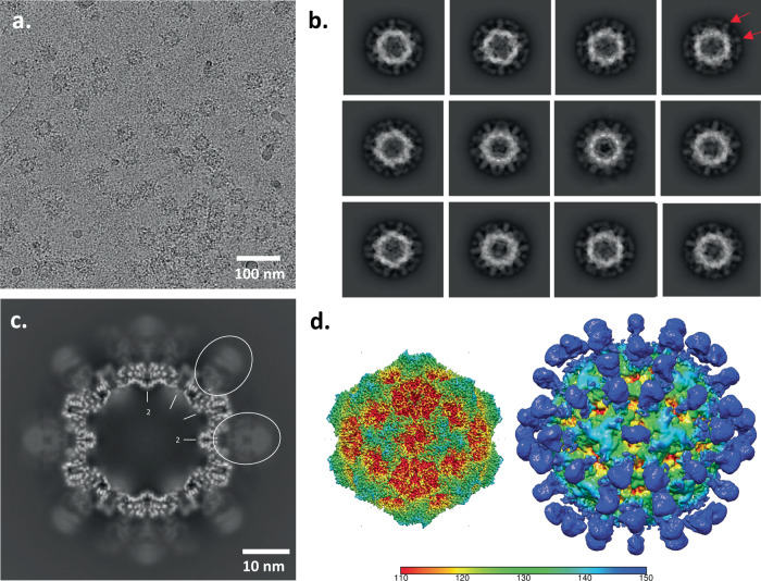
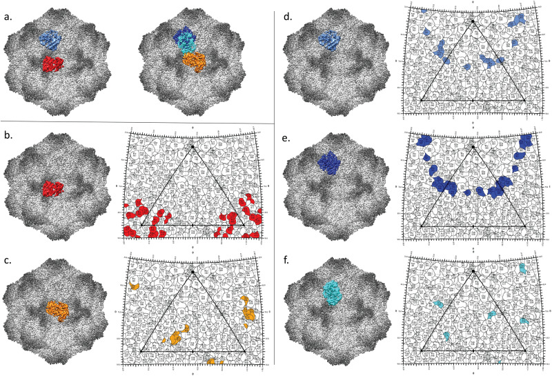
Canine parvovirus (CPV) is an important pathogen that emerged by cross-species transmission to cause severe disease in dogs. To understand the host immune response to vaccination, sera from dogs immunized with parvovirus are obtained, the polyclonal antibodies are purified and used to solve the high resolution cryo EM structures of the polyclonal Fab-virus complexes. We use a custom software, Icosahedral Subparticle Extraction and Correlated Classification (ISECC) to perform subparticle analysis and reconstruct polyclonal Fab-virus complexes from two different dogs eight and twelve weeks post vaccination. In the resulting polyclonal Fab-virus complexes there are a total of five distinct Fabs identified. In both cases, any of the five antibodies identified would interfere with receptor binding. This polyclonal mapping approach identifies a specific, limited immune response to the live vaccine virus and allows us to investigate the binding of multiple different antibodies or ligands to virus capsids.
© 2023. Springer Nature Limited.
Conflict of interest statement
The authors declare no competing interests.
Figures


Similar articles
-
High-resolution asymmetric structure of a Fab-virus complex reveals overlap with the receptor binding site.Proc Natl Acad Sci U S A. 2021 Jun 8;118(23):e2025452118. doi: 10.1073/pnas.2025452118. Proc Natl Acad Sci U S A. 2021. PMID: 34074770 Free PMC article.
-
Near-Atomic Resolution Structure of a Highly Neutralizing Fab Bound to Canine Parvovirus.J Virol. 2016 Oct 14;90(21):9733-9742. doi: 10.1128/JVI.01112-16. Print 2016 Nov 1. J Virol. 2016. PMID: 27535057 Free PMC article.
-
Prevalence of positive antibody test results for canine parvovirus (CPV) and canine distemper virus (CDV) and response to modified live vaccination against CPV and CDV in dogs entering animal shelters.Vet Microbiol. 2012 May 25;157(1-2):86-90. doi: 10.1016/j.vetmic.2011.12.030. Epub 2011 Dec 30. Vet Microbiol. 2012. PMID: 22261239
-
Evolution of canine parvovirus--a need for new vaccines?Vet Microbiol. 2006 Oct 5;117(1):9-13. doi: 10.1016/j.vetmic.2006.04.003. Epub 2006 Apr 18. Vet Microbiol. 2006. PMID: 16765539 Review.
-
[Prevention of canine parvovirosis - Part 1: Humoral and cellular immunity].Tierarztl Prax Ausg K Kleintiere Heimtiere. 2021 Feb;49(1):44-50. doi: 10.1055/a-1319-4564. Epub 2021 Feb 15. Tierarztl Prax Ausg K Kleintiere Heimtiere. 2021. PMID: 33588464 Review. German.
Cited by
-
Overview of Recent Advances in Canine Parvovirus Research: Current Status and Future Perspectives.Microorganisms. 2024 Dec 30;13(1):47. doi: 10.3390/microorganisms13010047. Microorganisms. 2024. PMID: 39858815 Free PMC article. Review.
-
Structures and functions of the limited natural polyclonal antibody response to parvovirus infection.Proc Natl Acad Sci U S A. 2025 Feb 25;122(8):e2423460122. doi: 10.1073/pnas.2423460122. Epub 2025 Feb 14. Proc Natl Acad Sci U S A. 2025. PMID: 39951487 Free PMC article.
-
Current Review of Monoclonal Antibody Therapeutics in Small Animal Medicine.Animals (Basel). 2025 Feb 7;15(4):472. doi: 10.3390/ani15040472. Animals (Basel). 2025. PMID: 40002954 Free PMC article. Review.
-
Distinct evolutionary patterns of endemic and emerging parvoviruses, and the origin of a new pandemic virus.bioRxiv [Preprint]. 2025 Jul 18:2025.07.14.664739. doi: 10.1101/2025.07.14.664739. bioRxiv. 2025. PMID: 40791396 Free PMC article. Preprint.
References
Publication types
MeSH terms
Substances
Grants and funding
LinkOut - more resources
Full Text Sources
