

Vaginal Bacteria Elicit Acute Inflammatory Response in Fallopian Tube Organoids
- PMID: 37726587
- PMCID: PMC11378751
- DOI: 10.1007/s43032-023-01350-5
Vaginal Bacteria Elicit Acute Inflammatory Response in Fallopian Tube Organoids
Abstract
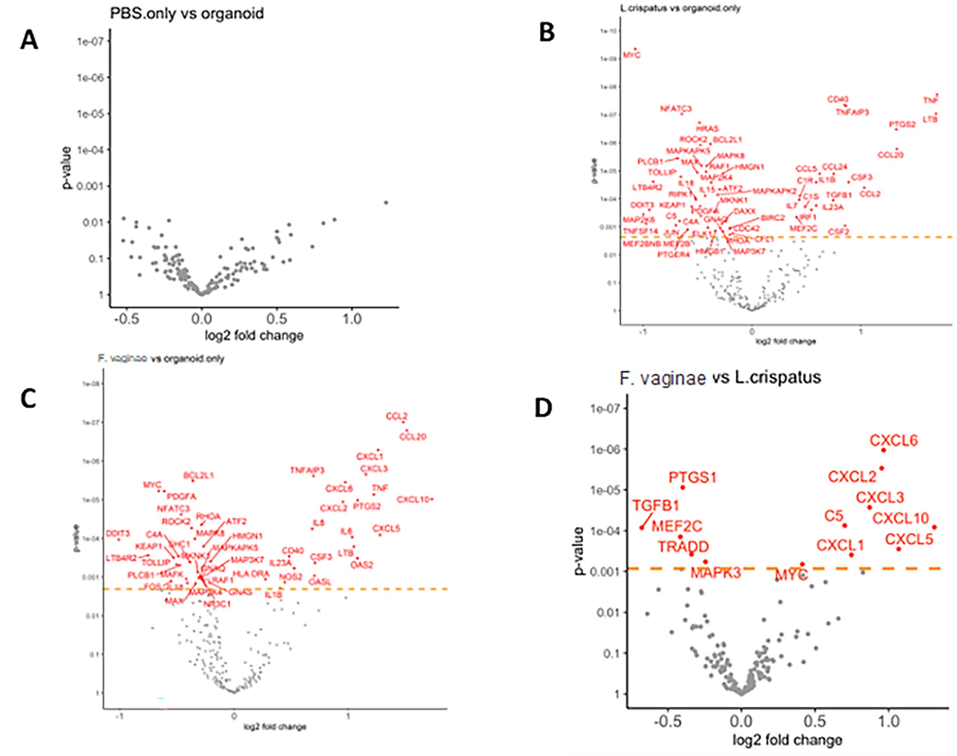
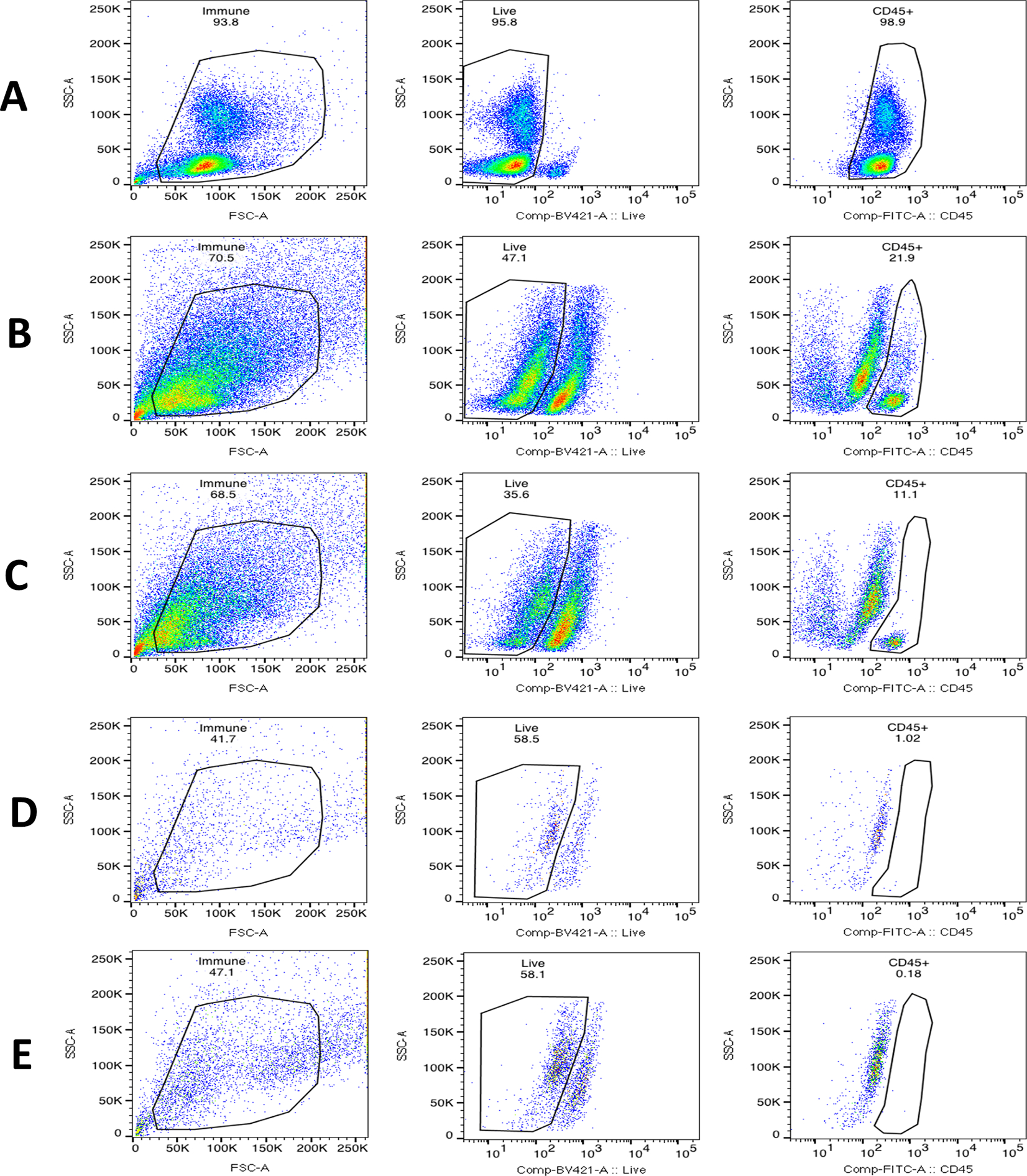
To facilitate in vitro mechanistic studies in pelvic inflammatory disease and subsequent tubal factor infertility, we sought to establish patient tissue derived fallopian tube (FT) organoids and to study their inflammatory response to acute vaginal bacterial infection. FT tissues were obtained from four patients after salpingectomy for benign gynecological diseases. We introduced acute infection in the FT organoid culture system by inoculating the organoid culture media with two common vaginal bacterial species, Lactobacillus crispatus and Fannyhessea vaginae. The inflammatory response elicited in the organoids after acute bacterial infection was analyzed by the expression profile of 249 inflammatory genes. Compared to the negative controls that were not cultured with any bacteria, the organoids cultured with either bacterial species showed multiple differentially expressed inflammatory genes. Marked differences were noted between the Lactobacillus crispatus infected organoids and those infected by Fannyhessea vaginae. Genes from the C-X-C motif chemokine ligand (CXCL) family were highly upregulated in Fannyhessea vaginae infected organoids. Flow cytometry showed that immune cells quickly disappeared during the organoid culture, indicating the inflammatory response observed with bacterial culture was generated by the epithelial cells in the organoids. In summary, we have shown that patient tissue derived FT organoids respond to acute bacterial infection with upregulation of inflammatory genes specific to different vaginal bacterial species. FT organoids is a useful in vitro model system to study the host-pathogen interaction during bacterial infection.
Keywords: Fallopian tube organoids; Host-pathogen interaction; Pelvic inflammatory disease (PID); Tubal factor infertility; Vaginal bacteria.
© 2023. The Author(s), under exclusive licence to Society for Reproductive Investigation.
Conflict of interest statement
Conflict of interest disclosures:
The authors declare that there are no conflicts of interests.
Figures


Update of
-
Vaginal bacteria elicit acute inflammatory response in fallopian tube organoids: a model for pelvic inflammatory disease.bioRxiv [Preprint]. 2023 Feb 7:2023.02.06.527402. doi: 10.1101/2023.02.06.527402. bioRxiv. 2023. Update in: Reprod Sci. 2024 Feb;31(2):505-513. doi: 10.1007/s43032-023-01350-5. PMID: 36798329 Free PMC article. Updated. Preprint.
-
Vaginal bacteria elicit acute inflammatory response in fallopian tube organoids: a model for pelvic inflammatory disease.Res Sq [Preprint]. 2023 May 23:rs.3.rs-2891189. doi: 10.21203/rs.3.rs-2891189/v1. Res Sq. 2023. Update in: Reprod Sci. 2024 Feb;31(2):505-513. doi: 10.1007/s43032-023-01350-5. PMID: 37293093 Free PMC article. Updated. Preprint.
Similar articles
-
Vaginal bacteria elicit acute inflammatory response in fallopian tube organoids: a model for pelvic inflammatory disease.bioRxiv [Preprint]. 2023 Feb 7:2023.02.06.527402. doi: 10.1101/2023.02.06.527402. bioRxiv. 2023. Update in: Reprod Sci. 2024 Feb;31(2):505-513. doi: 10.1007/s43032-023-01350-5. PMID: 36798329 Free PMC article. Updated. Preprint.
-
Vaginal bacteria elicit acute inflammatory response in fallopian tube organoids: a model for pelvic inflammatory disease.Res Sq [Preprint]. 2023 May 23:rs.3.rs-2891189. doi: 10.21203/rs.3.rs-2891189/v1. Res Sq. 2023. Update in: Reprod Sci. 2024 Feb;31(2):505-513. doi: 10.1007/s43032-023-01350-5. PMID: 37293093 Free PMC article. Updated. Preprint.
-
Protocol for the Detection of Organoid-Initiating Cell Activity in Patient-Derived Single Fallopian Tube Epithelial Cells.Methods Mol Biol. 2022;2429:445-454. doi: 10.1007/978-1-0716-1979-7_30. Methods Mol Biol. 2022. PMID: 35507180
-
Pathogenesis of Neisseria gonorrhoeae and the Host Defense in Ascending Infections of Human Fallopian Tube.Front Immunol. 2018 Nov 21;9:2710. doi: 10.3389/fimmu.2018.02710. eCollection 2018. Front Immunol. 2018. PMID: 30524442 Free PMC article. Review.
-
From mice to women and back again: causalities and clues for Chlamydia-induced tubal ectopic pregnancy.Fertil Steril. 2012 Nov;98(5):1175-85. doi: 10.1016/j.fertnstert.2012.07.1113. Epub 2012 Aug 9. Fertil Steril. 2012. PMID: 22884019 Review.
Cited by
-
Exploring the Relationship between Ovarian Cancer and Genital Microbiota: A Systematic Review and Meta-Analysis.J Pers Med. 2024 Mar 27;14(4):351. doi: 10.3390/jpm14040351. J Pers Med. 2024. PMID: 38672978 Free PMC article. Review.
-
Generation of healthy bovine ovarian organoids: a proof-of-concept derivation technique.J Ovarian Res. 2025 May 22;18(1):106. doi: 10.1186/s13048-025-01673-8. J Ovarian Res. 2025. PMID: 40405269 Free PMC article.
-
Addressing Key Questions in Organoid Models: Who, Where, How, and Why?Int J Mol Sci. 2023 Nov 6;24(21):16014. doi: 10.3390/ijms242116014. Int J Mol Sci. 2023. PMID: 37958996 Free PMC article. Review.
-
Advanced Technologies for Studying Microbiome-Female Reproductive Tract Interactions: Organoids, Organoids-on-a-Chip, and Beyond.Semin Reprod Med. 2023 Sep;41(5):160-171. doi: 10.1055/s-0043-1778067. Epub 2024 Jan 23. Semin Reprod Med. 2023. PMID: 38262440 Free PMC article. Review.
References
MeSH terms
Grants and funding
LinkOut - more resources
Full Text Sources
Medical
