. 2023 Sep 20;7(1):54.
doi: 10.1186/s41747-023-00369-2.

Radiomics-based prediction of FIGO grade for placenta accreta spectrum

Affiliations

Radiomics-based prediction of FIGO grade for placenta accreta spectrum

Helena C Bartels et al. Eur Radiol Exp. .

Erratum in

Abstract

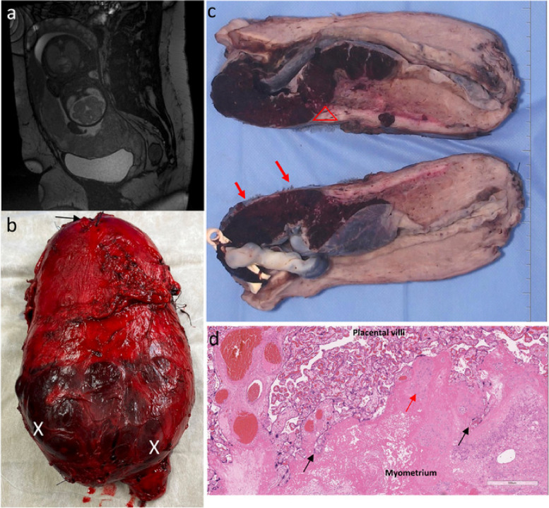
Background: Placenta accreta spectrum (PAS) is a rare, life-threatening complication of pregnancy. Predicting PAS severity is critical to individualise care planning for the birth. We aim to explore whether radiomic analysis of T2-weighted magnetic resonance imaging (MRI) can predict severe cases by distinguishing between histopathological subtypes antenatally.

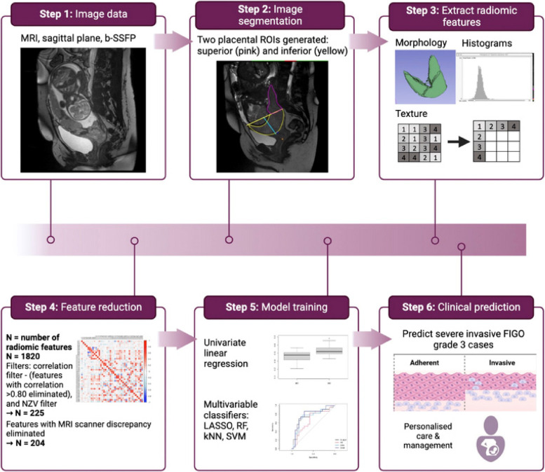
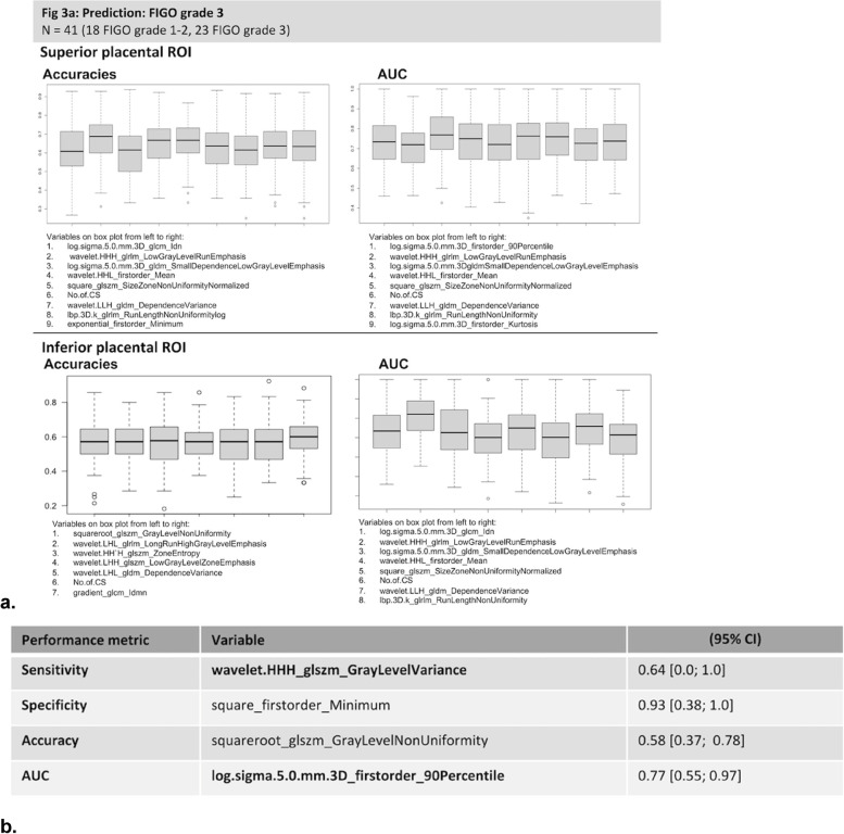
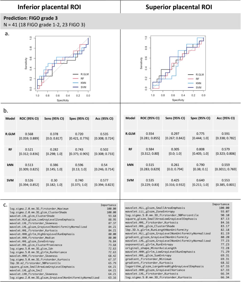
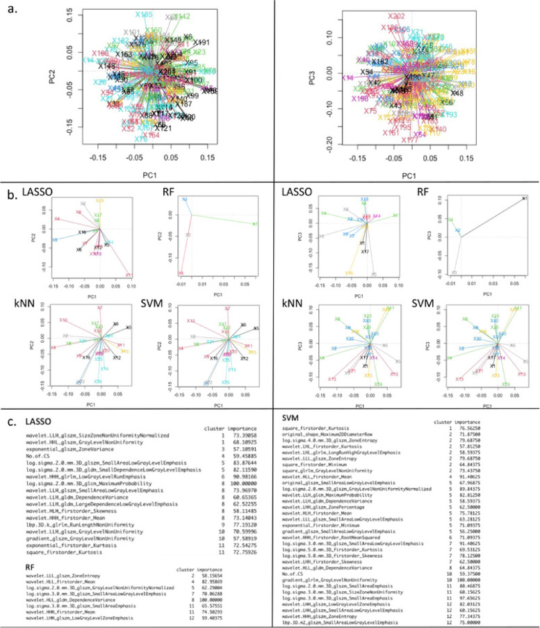
Methods: This was a bi-centre retrospective analysis of a prospective cohort study conducted between 2018 and 2022. Women who underwent MRI during pregnancy and had histological confirmation of PAS were included. Radiomic features were extracted from T2-weighted images. Univariate regression and multivariate analyses were performed to build predictive models to differentiate between non-invasive (International Federation of Gynecology and Obstetrics [FIGO] grade 1 or 2) and invasive (FIGO grade 3) PAS using R software. Prediction performance was assessed based on several metrics including sensitivity, specificity, accuracy and area under the curve (AUC) at receiver operating characteristic analysis.

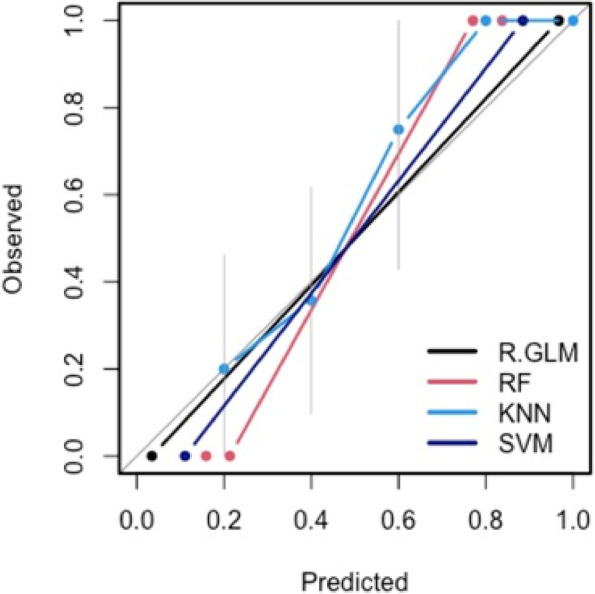
Results: Forty-one women met the inclusion criteria. At univariate analysis, 0.64 sensitivity (95% confidence interval [CI] 0.0-1.00), specificity 0.93 (0.38-1.0), 0.58 accuracy (0.37-0.78) and 0.77 AUC (0.56-.097) was achieved for predicting severe FIGO grade 3 PAS. Using a multivariate approach, a support vector machine model yielded 0.30 sensitivity (95% CI 0.18-1.0]), 0.74 specificity (0.38-1.00), 0.58 accuracy (0.40-0.82), and 0.53 AUC (0.40-0.85).

Conclusion: Our results demonstrate a predictive potential of this machine learning pipeline for classifying severe PAS cases.

Relevance statement: This study demonstrates the potential use of radiomics from MR images to identify severe cases of placenta accreta spectrum antenatally.

Key points: • Identifying severe cases of placenta accreta spectrum from imaging is challenging. • We present a methodological approach for radiomics-based prediction of placenta accreta. • We report certain radiomic features are able to predict severe PAS subtypes. • Identifying severe PAS subtypes ensures safe and individualised care planning for birth.

Keywords: Machine learning; Magnetic resonance imaging; Placenta accreta; Pregnancy; Radiomics.


Conflict of interest statement

Jim O’Doherty is an employee of Siemens Medical Solutions (which did not sponsor or fund this study). The remaining authors have no conflicts of interest to declare.

Figures

Fig. 1
Fig. 1
A case of PAS FIGO grade 3: correlation between MRI and histopathology. a MRI sagittal view obtained at 30 weeks gestation. Complete placenta previa demonstrating features of PAS including abnormal intraplacental vascularity, myometrial thinning and placental bulge towards the bladder. b Fresh hysterectomy specimen showing lower uterine segment bulging and distention with minimal overlying serosa (X) from abnormal placentation. Placenta can be seen through the very thin remaining serosa (X). Arrow marks fundal uterine incision where the baby was delivered. c Gross cross section of cut specimen: FIGO 3a with outer 25% of the myometrium involved. Triangle marks area where area of placental "invasion" led to scar dehiscence, with only a thin area of residual myometrium remaining (red arrows). No invasion beyond serosa or involvement of other organs. d Microscopy shows invaded placenta with absent decidua basalis, trophoblast cells invading deep into the myometrium (black arrows) as a result of abnormal uterine remodelling from a previous Caesarean scar, and loss of the normal uterine contour. Evidence of chronic inflammation (red arrow) and edema are also present in the myometrium. MRI Magnetic resonance imaging, FIGO International Federation of Gynecology and Obstetrics, PAS Placenta accreta spectrum
Fig. 2
Fig. 2
Summary of radiomics processing, feature reduction, and modelling. The N reported throughout applies to the number of radiomic features extracted when using convolutional image filters. kNN k-nearest neighbour, LASSO Least absolute shrinkage and selection operator, NZR Near zero variance, RF Random forest, SVM Support vector machine
Fig. 3
Fig. 3
Univariate bootstrapped linear regression models. a Box-plots showing performance metrics of models as estimated by accuracy, area under the curve (AUC) for top performing radiomic features. b Radiomic features with top performance metrics from univariate analysis; the table lists the variables with the highest performance for each performance metric of sensitivity, specificity, accuracy, and AUC from the univariate bootstrapped linear regression analysis (in bold, superior placental region of interest)
Fig. 4
Fig. 4
Model calibration curves with associated 95% confidence bands (grey). The y-axis represents the actual probability, and the x-axis represents the predicted probability of placenta accreta spectrum. Each curve corresponds to a predictive model and assesses the alignment between mean estimated model probabilities obtained from that model, and observed event rates within each risk group. Here the risk groups are defined with respect to the quintiles of the predicted probabilities from that model. The closer the lines are to the ideal grey line (45° line), the better the prediction accuracy of the model
Fig. 5
Fig. 5
Multivariate bootstrapped models. The performance of each model for predicting severe FIGO grade 3 PAS from the inferior and superior placental ROI is shown. In panels a and b, the ROC curve and performance metrics for each of the models for predicting invasive FIGO grade 3 PAS are shown. Panel c reports the variable importance for radiomic and clinical features used in the prediction for SVM from the inferior and superior placental ROIs. CS Caesarean section, FIGO International Federation of Gynecology and Obstetrics, kNN: k-nearest neighbour, PAS Placenta accreta spectrum, R-GLM LASSO, Least absolute shrinkage and selection operator, RF Random forest, ROI Region of interest, SVM Support vector machine
Fig. 6
Fig. 6
Principal component analysis (PCA) from superior placental region of interest using all radiomic features from image filters. Panel a shows how the feature space used by each model from whole dataset. The plots show each model is using information for making predictions from different areas of the feature space. Panel b shows PCA for each model. Panel c shows the radiomic features within each PCA cluster that were important for each prediction model. This suggests some radiomic features could be used either interchangeably or in combination for placenta accreta spectrum prediction. **kNN yielded variable importance and PCA outputs identical to those obtained from support vector machine (SVM) as seen in panel b (due to the discretisation method used in evaluating variable importance for these two models) and therefore only the output for SVM are shown in c

References

    1. Bartels HC, Postle JD, Downey P, Brennan DJ. Placenta accreta spectrum: a review of pathology, molecular biology, and biomarkers. Dis Markers. 2018;2018:1507674. doi: 10.1155/2018/1507674. - DOI - PMC - PubMed
    1. Jauniaux E, Ayres-de-Campos D, Langhoff-Roos J, Fox KA, Collins S. FIGO classification for the clinical diagnosis of placenta accreta spectrum disorders. Int J Gynaecol Obstet. 2019;146:20–24. doi: 10.1002/ijgo.12761. - DOI - PubMed
    1. Bartels HC, Rogers AC, O'Brien D, McVey R, Walsh J, Brennan DJ. Association of implementing a multidisciplinary team approach in the management of morbidly adherent placenta with maternal morbidity and mortality. Obstet Gynecol. 2018;132:1167–1176. doi: 10.1097/aog.0000000000002865. - DOI - PubMed
    1. Einerson BD, Silver RM. Multidisciplinary teams in the management of placenta accreta spectrum disorders. Curr Obstet Gynecol Rep Rep. 2019;8:80–85. doi: 10.1007/s13669-019-00264-x. - DOI
    1. Jha P, Pōder L, Bourgioti C, et al. Society of Abdominal Radiology (SAR) and European Society of Urogenital Radiology (ESUR) joint consensus statement for MR imaging of placenta accreta spectrum disorders. Eur Radiol. 2020;30:2604–2615. doi: 10.1007/s00330-019-06617-7. - DOI - PubMed

Publication types

LinkOut - more resources