

Alterations in choroidal circulatory dynamics and choroidal thickness before and after treatment in posterior scleritis
- PMID: 37726746
- PMCID: PMC10508002
- DOI: 10.1186/s12886-023-03140-8
Alterations in choroidal circulatory dynamics and choroidal thickness before and after treatment in posterior scleritis
Abstract
Background: Posterior scleritis is an inflammatory reaction of the sclera that occurs posterior to the ora serrata. The aim of this study was to present a case of posterior scleritis and to analyze choroidal circulatory and structural changes using laser speckle flowgraphy (LSFG) and optical coherence tomography (OCT), respectively.
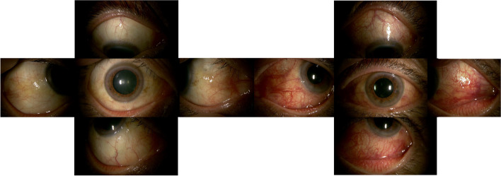
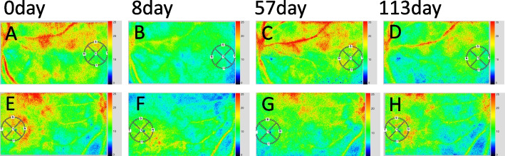
Case presentation: A 64-year-old man presented to our department because of hyperemia of the left eye for one week, diplopia, ocular pain, and distorted vision when looking leftward. At an initial examination, his best-corrected visual acuity was 1.0 Oculi uterque (OU), with mild conjunctival hyperemia oculus dexter (OD) and marked ciliary hyperemia oculus sinister (OS). Color fundus photographs revealed a cluster of choroidal folds extending from the macula to the inferior retinal region OS. Swept-Source OCT showed choroidal thickening OD, and bacillary layer detachment and paracentral middle maculopathy on the paracentral side of the optic nerve papilla, suggesting severe inflammation. Fluorescein angiography showed hyperfluorescence in the optic disc and window defects around the macula OU. Indocyanine green angiography showed mottled choroidal vascular hyperpermeability findings in the late stage. B-mode echography displayed thickening of the posterior wall of the left eye. Orbital magnetic resonance imaging showed the thickened posterior eyeball. The patient was diagnosed with posterior scleritis, and 30 mg of oral prednisolone was then given and tapered off over the next 4 months. The hyperemia and intraocular inflammation resolved after the treatment. The rate of change in macular blood flow assessed by the mean blur rate on LSFG was 20.5% and 20.2% decrease OD and OS, respectively, before and after treatment. The central choroidal thickness showed 8.8% and 37.8% decrease OD and OS, respectively.
Conclusion: Posterior scleritis complicated with choroiditis was suggested to show different choroidal circulatory dynamics from those in other choroidal inflammations.
Keywords: Choroidal circulatory dynamics; Choroiditis; Laser speckle flowgraphy; optical coherence tomography; Posterior scleritis.
© 2023. BioMed Central Ltd., part of Springer Nature.
Conflict of interest statement
The authors declare no competing interests.
Figures


Similar articles
-
Choroidal Circulatory and Vascular Morphological Changes in Acute Macular Neuroretinopathy After Infection With Severe Acute Respiratory Syndrome Coronavirus 2: A Case Report With Literature Review.In Vivo. 2023 Nov-Dec;37(6):2869-2876. doi: 10.21873/invivo.13405. In Vivo. 2023. PMID: 37905626 Free PMC article. Review.
-
A case of choroidal melanocytosis observed by multimodal imaging with laser speckle flowgraphy.BMC Ophthalmol. 2023 Apr 26;23(1):180. doi: 10.1186/s12886-023-02933-1. BMC Ophthalmol. 2023. PMID: 37101259 Free PMC article.
-
Alterations of choroidal circulation and choroidal thickness before and after chemoradiotherapy in a case of metastatic choroidal tumor.BMC Ophthalmol. 2023 Jun 13;23(1):270. doi: 10.1186/s12886-023-03026-9. BMC Ophthalmol. 2023. PMID: 37312082 Free PMC article.
-
Alterations of choroidal circulation and vascular morphology in a patient with chronic myeloid leukemia before and after chemotherapy.BMC Ophthalmol. 2022 Apr 7;22(1):160. doi: 10.1186/s12886-022-02380-4. BMC Ophthalmol. 2022. PMID: 35392846 Free PMC article.
-
Multimodal imaging in sclerochoroidal calcification: a case report and literature review.BMC Ophthalmol. 2020 Jun 22;20(1):248. doi: 10.1186/s12886-020-01520-y. BMC Ophthalmol. 2020. PMID: 32571266 Free PMC article. Review.
References
Publication types
MeSH terms
LinkOut - more resources
Full Text Sources
