. 2023 Sep 19;43(1):44.
doi: 10.1186/s41232-023-00293-3.

Effect of JAK inhibitors on the three forms of bone damage in autoimmune arthritis: joint erosion, periarticular osteopenia, and systemic bone loss

Affiliations

Effect of JAK inhibitors on the three forms of bone damage in autoimmune arthritis: joint erosion, periarticular osteopenia, and systemic bone loss

Masatsugu Komagamine et al. Inflamm Regen. .

Abstract

Background: The types of bone damage in rheumatoid arthritis (RA) include joint erosion, periarticular osteoporosis, and systemic osteoporosis. Janus kinase (JAK) inhibitors ameliorate inflammation and joint erosion in RA, but their effect on the three types of bone loss have not been reportedly explored in depth. We aimed to clarify how JAK inhibitors influence the various types of bone loss in arthritis by modulating osteoclastic bone resorption and/or osteoblastic bone formation.

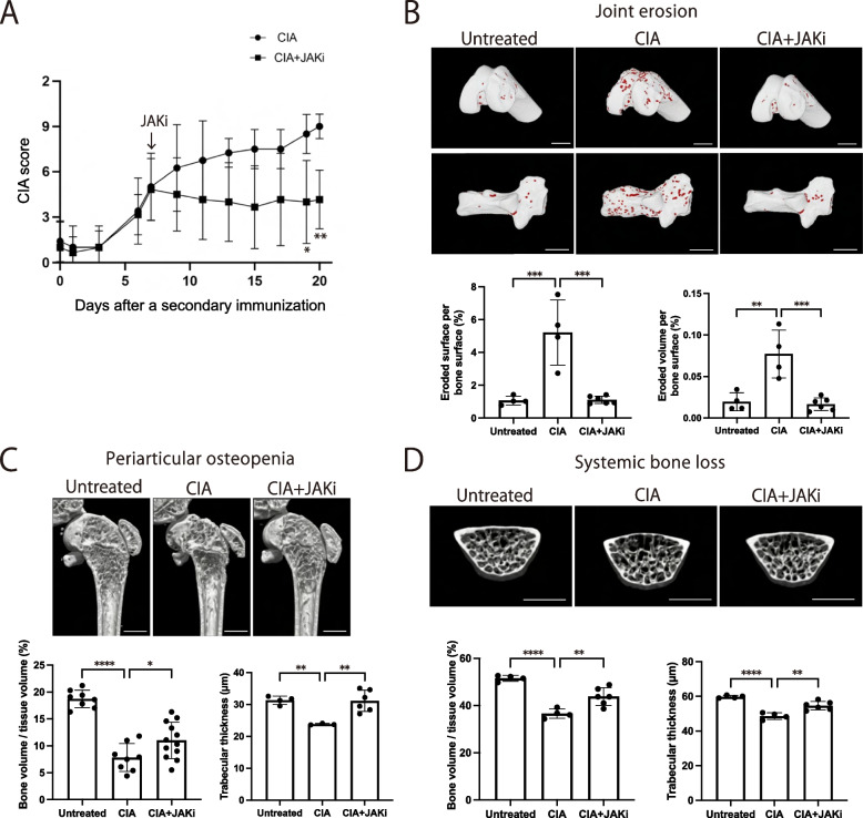
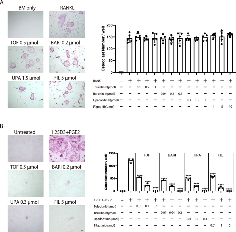
Methods: Collagen-induced arthritis (CIA) mice were treated with a JAK inhibitor after the onset of arthritis. Micro-computed tomography (μCT) and histological analyses (bone morphometric analyses) on the erosive calcaneocuboid joint, periarticular bone (distal femur or proximal tibia), and vertebrae were performed. The effect of four different JAK inhibitors on osteoclastogenesis under various conditions was examined in vitro.

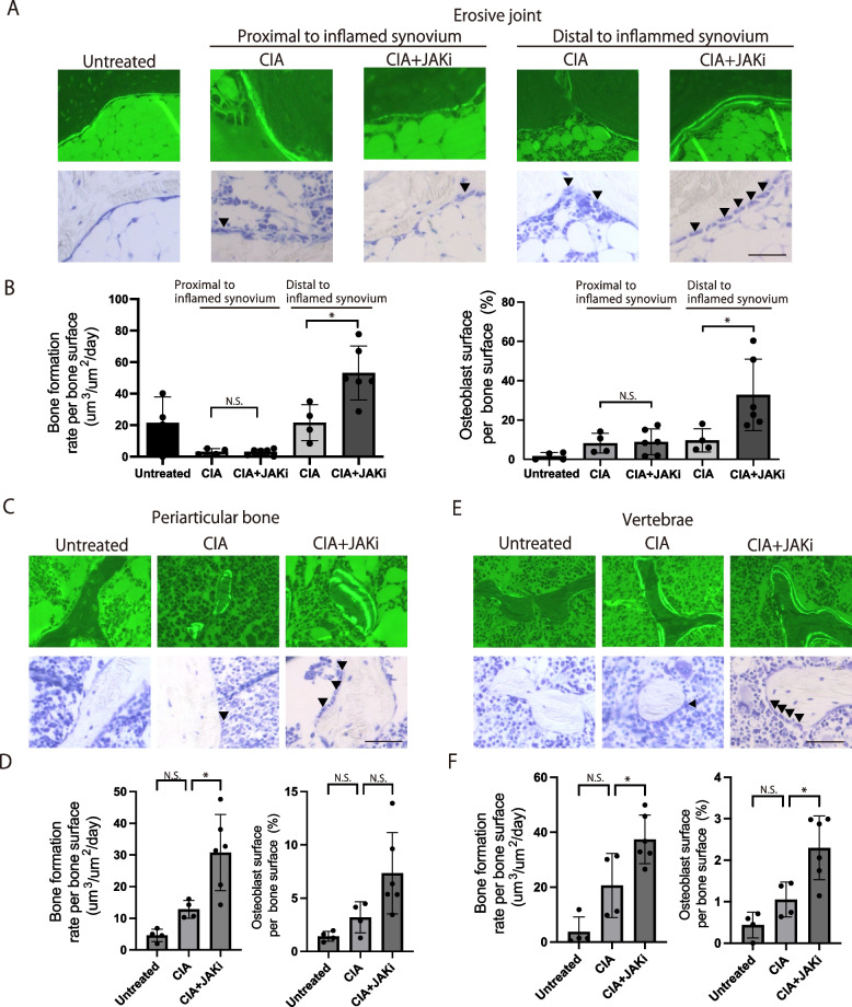
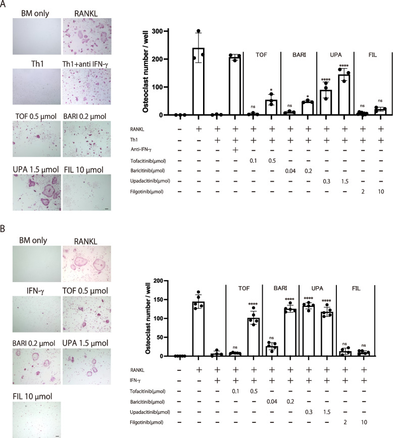
Results: The JAK inhibitor ameliorated joint erosion, periarticular osteopenia and systemic bone loss. It reduced the osteoclast number in all the three types of bone damage. The JAK inhibitor enhanced osteoblastic bone formation in the calcaneus distal to inflammatory synovium in the calcaneocuboid joints, periarticular region of the tibia and vertebrae, but not the inflamed calcaneocuboid joint. All the JAK inhibitors suppressed osteoclastogenesis in vitro to a similar extent in the presence of osteoblastic cells. Most of the JAK inhibitors abrogated the suppressive effect of Th1 cells on osteoclastogenesis by inhibiting IFN-γ signaling in osteoclast precursor cells, while a JAK inhibitor did not affect this effect due to less ability to inhibit IFN-γ signaling.

Conclusions: The JAK inhibitor suppressed joint erosion mainly by inhibiting osteoclastogenesis, while it ameliorated periarticular osteopenia and systemic bone loss by both inhibiting osteoclastogenesis and promoting osteoblastogenesis. These results indicate that the effect of JAK inhibitors on osteoclastogenesis and osteoblastogenesis depends on the bone damage type and the affected bone area. In vitro studies suggest that while JAK inhibitors inhibit osteoclastic bone resorption, their effects on osteoclastogenesis in inflammatory environments vary depending on the cytokine milieu, JAK selectivity and cytokine signaling specificity. The findings reported here should contribute to the strategic use of antirheumatic drugs against structural damages in RA.

Keywords: Autoimmune arthritis; Bone damage; JAK inhibitor; Osteoblast; Osteoclast.


Conflict of interest statement

The Department of Osteoimmunology is an endowment department supported with an unrestricted grant from AYUMI Pharmaceutical Corporation, ELECOM, JCR Pharmaceuticals, Kondo Cotton Spinning, MIKIHOUSE, MITSUI FUDOSAN, Meiji, Noevir, TAKENAKA, TENNENBUTSU IKAGAKU KENKYU ZAIDAN and Yakult.

Figures

Fig. 1
Fig. 1
The effect of the JAK inhibitor on bone erosion, periarticular osteopenia and systemic bone loss in autoimmune arthritis. A Arthritis score of the CIA mice (n = 4) and CIA mice administered the JAK inhibitor upadacitinib (CIA + JAKi mice) (n = 6). The JAK inhibitor was administrated 1 week after the secondary immunization. B Representative μCT images (upper: knee joint, lower: calcaneocuboid joint), eroded surface (left) and eroded volume (right) per bone surface of the calcaneocuboid joint of untreated (n = 4), CIA (n = 4) and CIA + JAKi mice (n = 6). The red area indicates cavities. C Representative μCT images (upper panel), bone volume per tissue volume (BV/TV) (lower left) and trabecular thickness (lower right) of the distal femur of untreated (n = 4), CIA (n = 4), and CIA + JAKi mice (n = 6). D Representative μCT images (upper panel), BV/TV (lower left) and trabecular thickness (lower right) of lumbar vertebrae of untreated (n = 4), CIA (n = 4) and CIA + JAKi mice (n = 6). Scale bar: (1 mm) (B, C, D). All data are expressed as the mean ± SEM. *P < 0.05; **P < 0.01; ***P < 0.001; ****p < 0.0001; by unpaired Student’s t test (A), and one-way ANOVA with the Holm-Sidak multiple comparisons test (BD)
Fig. 2
Fig. 2
The effect of the JAK inhibitor on osteoclastogenic bone resorption in the three different types of bone damage in autoimmune arthritis. A, B Representative TRAP+ staining (A) and the number of TRAP+ multinucleated cells per bone surface (B) of the calcaneocuboid joint of untreated (n = 4), CIA (n = 4), and CIA + JAKi mice (n = 6). C, D Representative TRAP+ staining (C) and the number of TRAP+ multinucleated cells per bone surface (D) of periarticular bone (proximal tibia) of untreated (n = 4), CIA (n = 4) and CIA + JAKi mice (n = 6). E, F Representative TRAP+ staining (E) and the number of TRAP+ multinucleated cells per bone surface (F) of lumbar vertebrae of untreated (n = 4), CIA (n = 4), and CIA + JAKi mice (n = 6). Scale bar: (100 μm, upper left; 50 μm, others) (A, E). (100 μm, upper left; 25 μm, others) (C). All data are expressed as the mean ± SEM. *P < 0.05; **P < 0.01; ***P < 0.001; ****p < 0.0001; by one-way ANOVA with the Holm-Sidak multiple comparisons test (B, D, F)
Fig. 3
Fig. 3
The effect of the JAK inhibitor on osteoblastic bone formation in the three types of bone damage in autoimmune arthritis. A, B Representative calcein labeling (A, upper), toluidine blue staining (A, lower), bone formation rate (B, left) and osteoblast surface per bone surface of the calcaneus of untreated (n = 4), CIA (n = 4), and CIA + JAKi mice (n = 6). The calcaneus proximal and distal to inflamed synovium were investigated. C, D Representative calcein labeling (C, upper), toluidine blue staining (C, lower), bone formation rate (D, left), and osteoblast surface per bone surface of the periarticular bone (proximal tibia, D, right) of untreated (n = 4), CIA (n = 4), and CIA + JAKi mice (n = 6). E, F Representative calcein labeling (E, upper), toluidine blue staining (F, lower), bone formation rate (F, left) and osteoblast surface per bone surface of the lumbar vertebrae (F, right) of untreated (n = 4), CIA (n = 4), and CIA + JAKi mice (n = 6). Scale bar: (50 μm) (A, C, E). The arrowheads show osteoblasts. All data are expressed as the mean ± SEM. *P < 0.05; by one-way ANOVA with the Holm-Sidak multiple comparisons test (B, D, F) N.S., not significant
Fig. 4
Fig. 4
The effects of the various JAK inhibitors on osteoclastogenesis in a monoculture of osteoclast precursor cells and coculture system of precursors with supporting cells. A Representative TRAP staining (left) and number of TRAP+ multinucleated cells (right) in the presence of the respective JAK inhibitor in the monoculture of osteoclast precursors. B Representative TRAP staining (left) and number of TRAP+ multinucleated cells (right) in the presence of the respective JAK inhibitor in the coculture of osteoclast precursors and osteoblastic cells. Scale bar: (100 μm). All data are expressed as the mean ± SEM. ****p < 0.0001; by one-way ANOVA with the Holm-Sidak multiple comparisons test
Fig. 5
Fig. 5
The effects of the various JAK inhibitors on osteoclastogenesis in a RANKL-induced osteoclast formation system cocultured with Th1 cells. A Representative TRAP staining (left) and number of TRAP+ multinucleated cells (right) in the presence of the respective JAK inhibitor in the RANKL-induced osteoclast formation system cocultured with Th1 cells. B Representative TRAP staining (left) and number of TRAP+ multinucleated cells (right) in the presence of the respective JAK inhibitor and IFN-γ in the RANKL-induced osteoclast formation system. Scale bar: (100 µm). All data are expressed as the mean ± SEM. *P < 0.05, ****p < 0.0001; by one-way ANOVA with the Holm-Sidak’s multiple comparisons test

Similar articles

Cited by

References

    1. Firestein GS. Evolving concepts of rheumatoid arthritis. Nature. 2003;423:356–361. doi: 10.1038/nature01661. - DOI - PubMed
    1. McInnes IB, Schett G. The pathogenesis of rheumatoid arthritis. N Engl J Med. 2011;365:2205–2219. doi: 10.1056/NEJMra1004965. - DOI - PubMed
    1. Komatsu N, Takayanagi H. Mechanisms of joint destruction in rheumatoid arthritis - immune cell-fibroblast-bone interactions. Nat Rev Rheumatol. 2022;18:415–429. doi: 10.1038/s41584-022-00793-5. - DOI - PubMed
    1. Croft AP, Campos J, Jansen K, et al. Distinct fibroblast subsets drive inflammation and damage in arthritis. Nature. 2019;570:246–251.R. doi: 10.1038/s41586-019-1263-7. - DOI - PMC - PubMed
    1. Danks L, Komatsu N, Guerrini MM, et al. RANKL expressed on synovial fibroblasts is primarily responsible for bone erosions during joint inflammation. Ann Rheum Dis. 2016;75:1187–1195. doi: 10.1136/annrheumdis-2014-207137. - DOI - PubMed

LinkOut - more resources