

Magnetic resonance imaging findings of pulmonary sclerosing pneumocytoma: a case report and literature review
- PMID: 37727218
- PMCID: PMC10505615
- DOI: 10.3389/fonc.2023.1158328
Magnetic resonance imaging findings of pulmonary sclerosing pneumocytoma: a case report and literature review
Abstract
Background: Pulmonary sclerosing pneumocytoma (PSP) is a rare lung tumor that is mostly isolated and commonly reported among middle-aged East Asian women. Recently, Immunohistochemistry (IHC) analysis has suggested that PSP is of primitive epithelial origin, most likely derived from type II alveolar air cells. Patients with PSP are generally asymptomatic and usually detected for other unrelated reasons during routine imaging. Several studies have already investigated the computed tomography (CT) features of PSP and their correlation with pathology. Magnetic resonance imaging (MRI) is a radiation-free imaging technique with important diagnostic value for specific pulmonary nodules. However, very few case reports or studies focus on the MRI findings of PSP.
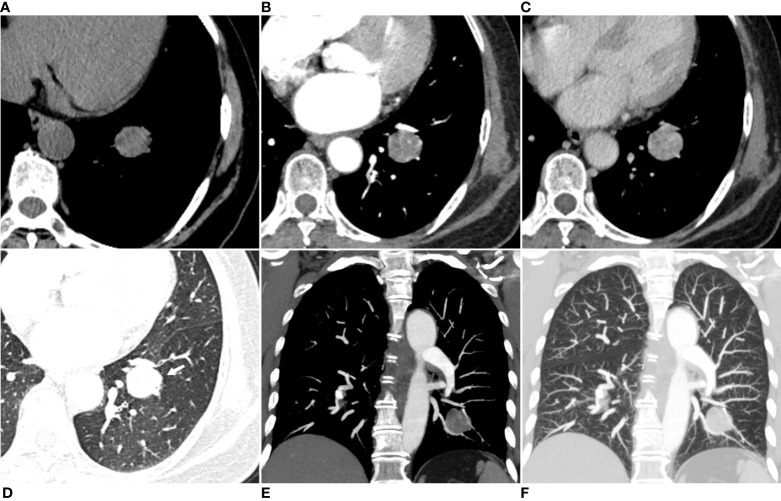
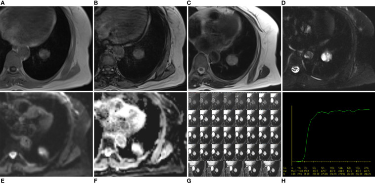
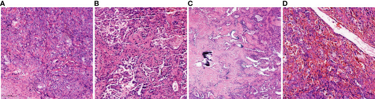
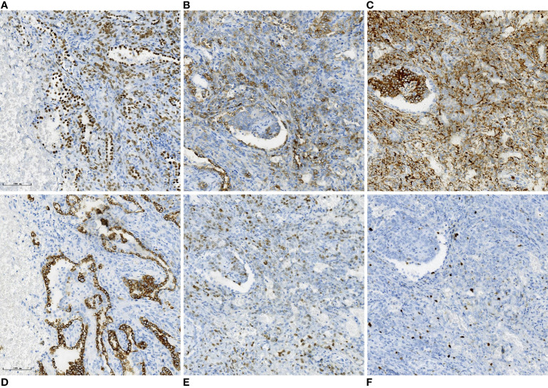
Case report: We reported a case of an asymptomatic 56-year-old female with a solitary, well-defined soft-tissue mass in the lower lobe of the left lung. The mass showed iso-to-high signal intensity (SI) than muscle on T1-weighted image (T1WI) and T2-weighted image (T2WI) and a much higher SI on fat-suppressed T2WI, diffusion-weighted image, and apparent diffusion coefficient image. Contrast-enhanced fat-suppressed T1WI revealed noticeable inhomogeneous progressive enhancement throughout the mass. The mass revealed early enhancement without a significant peak, followed by a plateau pattern on dynamic contrast-enhanced MRI images. The patient underwent left basal segmentectomy via thoracoscopic surgery. Histopathology and IHC results of the surgical specimen confirmed that it was a PSP. We concluded that the MRI findings of PSP might adequately reflect the different components within the tumor and aid clinicians in preoperative diagnosis and assessment. To the best of our knowledge, this is the most comprehensive case report on the MRI findings of PSP.
Conclusion: The MRI findings of PSP correspond to its histopathological features. Here, we present a case of PSP with the most comprehensive MRI findings, emphasizing the importance of multiple-sequence MRI in diagnosing PSP.
Keywords: case report; computed tomography; magnetic resonance imaging; pulmonary; pulmonary sclerosing pneumocytoma.
Copyright © 2023 Li, Yang, Gu, Wang, Deng, Feng, Zhang, Wang, Wang and Shi.
Conflict of interest statement
The authors declare that the research was conducted in the absence of any commercial or financial relationships that could be construed as a potential conflict of interest.
Figures
Similar articles
-
[Analysis of Clinical Characteristics of 35 Cases of Pulmonary Sclerosing Pneumocytoma].Zhongguo Fei Ai Za Zhi. 2020 Dec 20;23(12):1049-1058. doi: 10.3779/j.issn.1009-3419.2020.103.19. Zhongguo Fei Ai Za Zhi. 2020. PMID: 33357311 Free PMC article. Chinese.
-
Genome profile in a extremely rare case of pulmonary sclerosing pneumocytoma presenting with diffusely-scattered nodules in the right lung.Cancer Biol Ther. 2018 Jan 2;19(1):13-19. doi: 10.1080/15384047.2017.1360443. Epub 2017 Dec 22. Cancer Biol Ther. 2018. PMID: 29236566 Free PMC article.
-
A case report on incidentally detected pulmonary sclerosing pneumocytoma: a diagnostic challenge.Ann Med Surg (Lond). 2024 Aug 14;86(10):6194-6197. doi: 10.1097/MS9.0000000000002481. eCollection 2024 Oct. Ann Med Surg (Lond). 2024. PMID: 39359764 Free PMC article.
-
Pulmonary sclerosing pneumocytoma in an 18-year-old male patient: A case report and literature review.Medicine (Baltimore). 2020 Jun 26;99(26):e20869. doi: 10.1097/MD.0000000000020869. Medicine (Baltimore). 2020. PMID: 32590790 Free PMC article. Review.
-
Sclerosing Pneumocytoma with a Wax-and-Wane Pattern of Growth: A Case Report on Computed Tomography and Magnetic Resonance Imaging Findings and a Literature Review.Korean J Radiol. 2015 Jul-Aug;16(4):947-50. doi: 10.3348/kjr.2015.16.4.947. Epub 2015 Jul 1. Korean J Radiol. 2015. PMID: 26175598 Free PMC article. Review.
References
-
- Devouassoux-Shisheboran M, Hayashi T, Linnoila RI, Koss MN, Travis WD. A clinicopathologic study of 100 cases of pulmonary sclerosing hemangioma with immunohistochemical studies: TTF-1 is expressed in both round and surface cells, suggesting an origin from primitive respiratory epithelium. Am J Surg Pathol (2000) 24(7):906–16. doi: 10.1097/00000478-200007000-00002 - DOI - PubMed
Publication types
LinkOut - more resources
Full Text Sources
Miscellaneous