

Epithelioid Hemangioendothelioma of Tongue: A Rare Presentation
- PMID: 37727360
- PMCID: PMC10506149
- DOI: 10.30476/dentjods.2023.96208.1927
Epithelioid Hemangioendothelioma of Tongue: A Rare Presentation
Abstract
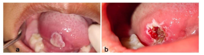
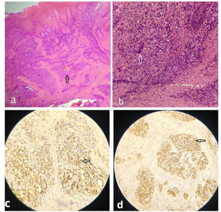
Hemangioendothelioma is a diverse set of proliferative and neoplastic vascular lesions with biological characteristics that fall halfway between benign hemangioma and malignant angiosarcoma. Hemangioendothelioma of the oral cavity is extremely rare and if present, it is seen on lips, gingiva, tongue, maxilla, and mandible. The following case report is about a lesion on the right ventrolateral border of the tongue of a six-year-old female patient. A Laser excision was done. Histopathology revealed the features of hemangioendothelioma. An immunohistochemical (IHC) study was done to correlate the findings with a histopathological picture and arrived at the final diagnosis of epithelioid hemangioendothelioma (EHE). The patient was followed up for two years and no recurrence was noticed.
Keywords: Epithelioid hemangioendothelium; Hemangioendothelioma; Immunohistochemistry; Tongue.
Copyright: © Journal of Dentistry.
Conflict of interest statement
The authors declare that they have no conflicts of interests.
Figures

Similar articles
-
Epithelioid Hemangioendothelioma of Mandibular Gingiva: A Challenging Diagnosis.J Clin Exp Dent. 2024 Sep 1;16(9):e1151-e1156. doi: 10.4317/jced.61925. eCollection 2024 Sep. J Clin Exp Dent. 2024. PMID: 39399857 Free PMC article.
-
Primary epithelioid angiosarcoma originating from the mandibular gingiva: a case report of an extremely rare oral lesion.World J Surg Oncol. 2020 Oct 3;18(1):260. doi: 10.1186/s12957-020-01999-1. World J Surg Oncol. 2020. PMID: 33010804 Free PMC article. Review.
-
Malignant pleuropulmonary epithelioid hemangioendothelioma - unusual presentation of an aggressive angiogenic neoplasm.Pathol Res Pract. 2014 Sep;210(9):613-8. doi: 10.1016/j.prp.2014.04.011. Epub 2014 May 27. Pathol Res Pract. 2014. PMID: 24939148
-
Ileal malignant hemangioendothelioma as a hypervascular lesion on computed tomography scan.Int J Surg Case Rep. 2014;5(1):19-21. doi: 10.1016/j.ijscr.2013.09.019. Epub 2013 Nov 26. Int J Surg Case Rep. 2014. PMID: 24394857 Free PMC article.
-
Primary malignant vascular tumors of the liver in children: Angiosarcoma and epithelioid hemangioendothelioma.World J Gastrointest Oncol. 2021 Apr 15;13(4):223-230. doi: 10.4251/wjgo.v13.i4.223. World J Gastrointest Oncol. 2021. PMID: 33889274 Free PMC article. Review.
Cited by
-
Immunohistochemical Analysis of Oral Spindle Cell Hemangioma.J Dent (Shiraz). 2025 Jun 1;26(2):194-198. doi: 10.30476/dentjods.2024.101499.2305. eCollection 2025 Jun. J Dent (Shiraz). 2025. PMID: 40510225 Free PMC article.
References
-
- Weiss SW, Enzinger FM. Epithelioid Hemangioendothelioma: a vascular tumor often mistaken for a carcinoma. Cancer. 1982; 50: 970–981. - PubMed
-
- Weiss SW, Goldblum JR. Haemangioendothelioma‑Vascular tumour of intermediate malignancy. In: Schmitt W, Black S, editors. Enzinger and Weiss’s Soft Tissue Tumours. 5th ed. Philadelphia: USA, Mosby Elsevier; 2008. pp. 681–687.
-
- Galateau-Salle F, Churg A, Roggli V, Travis W. The 2015 world health organization classification of tumors of the pleura: advances since the 2004 classification. J Thorac Oncol. 2016; 11: 142–154. - PubMed
Publication types
LinkOut - more resources
Full Text Sources