

Fraxin in Combination with Dexamethasone Attenuates LPS-Induced Liver and Heart Injury and Their Anticytokine Activity in Mice
- PMID: 37727368
- PMCID: PMC10506875
- DOI: 10.1155/2023/5536933
Fraxin in Combination with Dexamethasone Attenuates LPS-Induced Liver and Heart Injury and Their Anticytokine Activity in Mice
Abstract
Background: Cytokine storm syndrome (CSS) is a major cause of morbidity and mortality in people suffering from hyperinflammatory status, which diverse etiological factors, including pathogens, therapeutic interventions, malignancies, and autoimmune disorders, can instigate. Since there is limited research on the antioxidant properties of fraxin and no studies have investigated its potential as an anticytokine storm agent, it is important to note that most studies have primarily focused on proinflammatory cytokines such as IL-1β, IL-6, and TNFα during cytokine storm. However, little research discusses the role of chemokines, particularly IL-8, during cytokine storms. Therefore, further investigation is warranted into the role of fraxin as an anticytokine storm agent and the involvement of IL-8 in cytokine storms. The present study examines the preventive efficacy of fraxin and the combination of fraxin and dexamethasone (FD) in mitigating lipopolysaccharide-induced systemic inflammation in mice caused by Escherichia coli, 055: B5.
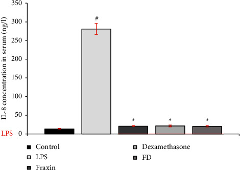
Methods: Five groups of ten mice were randomly assigned: LPS only group (5 mg/kg, intraperitoneally i.p.), control (normal saline N.S. 1 ml/kg, i.p.), concentrations were selected based on previous literature, fraxin (120 mg/kg, i.p.), dexamethasone (5 mg/kg, i.p.), fraxin + dexamethasone (FD) (60 mg/kg + 2.5 mg/kg, i.p.), administered one hour before LPS injection (5 mg/kg,i.p.), animals were euthanized next day, and interleukin-8 (IL-8) was quantified in serum using an enzyme-linked immunosorbent assay. The liver and heart tissues underwent histopathological analysis to assess morphological changes. For data analysis using ANOVA and Tukey post hoc tests, the Kruskal-Wallis and Mann-Whitney U tests were employed to analyze the histological results.
Results: A significant decline in IL-8 levels was recorded in the treatment groups almost to the same degree (p < 0.001), and the percentage of inhibition of IL-8 for fraxin, dexamethasone, and FD was 93%.92.4%, and 93%, respectively, compared to the LPS-only group. Histopathological scores were significantly reduced in liver and heart tissue (P < 0.05).
Conclusions: All interventions used in this study significantly reduced interleukin-8 (IL-8) levels and reduced LPS-induced liver and cardiac damage. The combination (FD) did not result in an evident superiority of either agent. More research is required to identify the possible usefulness of these agents in treating hyperinflammatory diseases, such as cytokine storms, in future clinical practice.
Copyright © 2023 Nada Sahib Shaker and Hayder Bahaa Sahib.
Conflict of interest statement
The authors declare that they have no conflicts of interest.
Figures


Similar articles
-
Anti-cytokine Storm Activity of Fraxin, Quercetin, and their Combination on Lipopolysaccharide-Induced Cytokine Storm in Mice: Implications in COVID-19.Iran J Med Sci. 2024 May 1;49(5):322-331. doi: 10.30476/ijms.2023.98947.3102. eCollection 2024 May. Iran J Med Sci. 2024. PMID: 38751871 Free PMC article.
-
Fraxin ameliorates lipopolysaccharide-induced acute lung injury in mice by inhibiting the NF-κB and NLRP3 signalling pathways.Int Immunopharmacol. 2019 Feb;67:1-12. doi: 10.1016/j.intimp.2018.12.003. Epub 2018 Dec 6. Int Immunopharmacol. 2019. PMID: 30530164
-
Fraxin inhibits lipopolysaccharide-induced inflammatory cytokines and protects against endotoxic shock in mice.Fundam Clin Pharmacol. 2020 Feb;34(1):91-101. doi: 10.1111/fcp.12500. Epub 2019 Aug 28. Fundam Clin Pharmacol. 2020. PMID: 31325387
-
Fraxin Alleviates LPS-Induced ARDS by Downregulating Inflammatory Responses and Oxidative Damages and Reducing Pulmonary Vascular Permeability.Inflammation. 2019 Oct;42(5):1901-1912. doi: 10.1007/s10753-019-01052-8. Inflammation. 2019. PMID: 31273573
-
Anticytokine therapies for acute inflammation and the systemic inflammatory response syndrome: IL-10 and ischemia/reperfusion injury as a new paradigm.Shock. 2000 Jun;13(6):425-34. doi: 10.1097/00024382-200006000-00002. Shock. 2000. PMID: 10847628 Review.
References
-
- Kopp F., Kupsch S., Schromm A. B. Lipopolysaccharide- binding protein is bound and internalized by host cells and colocalizes with LPS in the cytoplasm: implications for a role of LBP in intracellular LPS-signaling. Biochimica et Biophysica Acta (BBA)-Molecular Cell Research . 2016;1863(4):660–672. doi: 10.1016/j.bbamcr.2016.01.015. - DOI - PubMed
LinkOut - more resources
Full Text Sources
Miscellaneous
