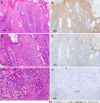
Malignant form of hidroacanthoma simplex: A case report
- PMID: 37727732
- PMCID: PMC10506014
- DOI: 10.12998/wjcc.v11.i24.5804
Malignant form of hidroacanthoma simplex: A case report
Abstract
Background: This paper presents a case of malignant hidroacanthoma simplex (HAS) and review the literature of previous cases to summarize the histopathological and immunohistochemical features and display the dermoscopic features of malignant HAS.
Case summary: We present an 88-year-old Asian female with malignant HAS. The diagnosis was made according to the histopathological and immunohistochemical results after biopsy. Previous case reports of malignant HAS were retrieved from PubMed to characterize the histopathological and immunohistochemical features. We also display the dermoscopic features of malignant HAS that have not been reported.
Conclusion: Our findings demonstrate that prompt surgical treatment is an effective strategy for malignant HAS. Histopathology and immunohistochemistry are valuable diagnostic tools. This is the first case report to display the dermoscopic features of malignant HAS, and we speculate that dermoscopy may contribute to the diagnosis of malignant HAS.
Keywords: Case report; Dermoscopy; Diagnosis; Histopathology; Immunohistochemistry; Malignant hidroacanthoma simplex.
©The Author(s) 2023. Published by Baishideng Publishing Group Inc. All rights reserved.
Conflict of interest statement
Conflict-of-interest statement: All the authors declare that they have no conflicts of interest to disclose.
Figures


Similar articles
-
Dermoscopic-Histopathological Correlation of Eccrine Poroma: An Observational Study.Dermatol Pract Concept. 2019 Oct 31;9(4):283-291. doi: 10.5826/dpc.0904a07. eCollection 2019 Oct. Dermatol Pract Concept. 2019. PMID: 31723462 Free PMC article.
-
Clinical, dermoscopic, and histopathologic findings of hidroacanthoma simplex: a case report and literature review.Dermatol Reports. 2022 Oct 27;15(2):9571. doi: 10.4081/dr.2022.9571. eCollection 2023 Jun 7. Dermatol Reports. 2022. PMID: 37426364 Free PMC article.
-
Dermoscopic features of hidroacanthoma simplex: Usefulness in distinguishing it from Bowen's disease and seborrheic keratosis.J Dermatol. 2015 Oct;42(10):1002-5. doi: 10.1111/1346-8138.12945. Epub 2015 May 19. J Dermatol. 2015. PMID: 25989988
-
Malignant hidroacanthoma simplex. A case report and literature review.Dermatology. 1995;190(1):72-6. doi: 10.1159/000246640. Dermatology. 1995. PMID: 7894103 Review.
-
Role of In Vivo Reflectance Confocal Microscopy in the Analysis of Melanocytic Lesions.Acta Dermatovenerol Croat. 2018 Apr;26(1):64-67. Acta Dermatovenerol Croat. 2018. PMID: 29782304 Review.
References
-
- Coburn JG, Smith JL. Hidroacanthoma simplex; an assessment of a selected group of intraepidermal basal cell epitheliomata and of their malignant homologues. Br J Dermatol. 1956;68:400–418. - PubMed
-
- Sun Kim M, Rae Lee H, Lee JH, Son SJ, Song KY. Malignant hidroacanthoma simplex: arising in hidroacanthoma simplex mimicking clonal seborrheic keratosis. Int J Dermatol. 2013;52:258–260. - PubMed
-
- Lee JY, Lin MH. Pigmented malignant hidroacanthoma simplex mimicking irritated seborrheic keratosis. J Cutan Pathol. 2006;33:705–708. - PubMed
-
- Bardach H. Hidroacanthoma simplex with in situ porocarcinoma. A case suggesting malignant transformation. J Cutan Pathol. 1978;5:236–248. - PubMed
-
- Kohli N, Kim SS, Jiang SI. Malignant hidroacanthoma simplex treated with Mohs surgery. Dermatol Surg. 2015;41:518–520. - PubMed
Publication types
LinkOut - more resources
Full Text Sources

