. 2023 Oct;16(10):e012018.
doi: 10.1161/CIRCEP.123.012018. Epub 2023 Sep 20.

Reversible Pulsed Electrical Fields as an In Vivo Tool to Study Cardiac Electrophysiology: The Advent of Pulsed Field Mapping

Affiliations

Reversible Pulsed Electrical Fields as an In Vivo Tool to Study Cardiac Electrophysiology: The Advent of Pulsed Field Mapping

Jacob S Koruth et al. Circ Arrhythm Electrophysiol. 2023 Oct.

Abstract

Background: During electrophysiological mapping of tachycardias, putative target sites are often only truly confirmed to be vital after observing the effect of ablation. This lack of mapping specificity potentiates inadvertent ablation of innocent cardiac tissue not relevant to the arrhythmia. But if myocardial excitability could be transiently suppressed at critical regions, their suitability as targets could be conclusively determined before delivering tissue-destructive ablation lesions. We studied whether reversible pulsed electric fields (PFREV) could transiently suppress electrical conduction, thereby providing a means to dissect tachycardia circuits in vivo.

Methods: PFREV energy was delivered from a 9-mm lattice-tip catheter to the atria of 12 swine and 9 patients, followed by serial electrogram assessments. The effects on electrical conduction were explored in 5 additional animals by applying PFREV to the atrioventricular node: 17 low-dose (PFREV-LOW) and 10 high-dose (PFREV-HIGH) applications. Finally, in 3 patients manifesting spontaneous tachycardias, PFREV was applied at putative critical sites.

Results: In animals, the immediate post-PFREV electrogram amplitudes diminished by 74%, followed by 78% recovery by 5 minutes. Similarly, in patients, a 69.9% amplitude reduction was followed by 84% recovery by 3 minutes. Histology revealed only minimal to no focal, superficial fibrosis. PFREV-LOW at the atrioventricular node resulted in transient PR prolongation and transient AV block in 59% and 6%, while PFREV-HIGH caused transient PR prolongation and transient AV block in 30% and 50%, respectively. The 3 tachycardia patients had atypical atrial flutters (n=2) and atrioventricular nodal reentrant tachycardia. PFREV at putative critical sites reproducibly terminated the tachycardias; ablation rendered the tachycardias noninducible and without recurrence during 1-year follow-up.

Conclusions: Reversible electroporation pulses can be applied to myocardial tissue to transiently block electrical conduction. This technique of pulsed field mapping may represent a novel electrophysiological tool to help identify the critical isthmus of tachycardia circuits.

Keywords: catheters; electroporation; follow-up studies; swine; transients and migrants.


Conflict of interest statement

Disclosures Dr Reddy: Affera-Medtronic: consultant, equity (stock); other disclosures not related to this article are detailed in the Supplemental Material. Dr Koruth: Affera-Medtronic: grant support, stock, consultant. Dr Neuzil: Affera-Medtronic: grant support.

Figures

Figure 1.
Figure 1.
Effect of reversible pulsed electric field (PFREV) on porcine atrial myocardium. A, A single PFREV application was placed between 2 discrete radiofrequency (RF) ablation lesions to facilitate identification of the PFREV lesion in the chronic phase. B, The PFREV lesion was identified as preserved voltage area in between RF scar on 3D voltage map. C, Histology demonstrated normal myocardium with no evidence of lesion formation between the 2 RF lesions.
Figure 2.
Figure 2.
Effect of reversible pulsed electric field (PFREV) on human atrial myocardium. Detailed high-density electroanatomic maps were created at baseline, immediately after, and at 1 minute post-PFREV to display local bipolar voltage change. Top uses the bipolar voltage range of 0.10 to 0.50 mV, and bottom uses the bipolar voltage range of 0.10 to 1.00 mV.
Figure 3.
Figure 3.
Atrial electrogram amplitude changes after reversible pulsed electric field (PFREV). A, Shown is an example of the acutely diminished electrograms observed on the mini-electrodes of the lattice catheter immediately after a PFREV application (red arrow). Note the immediately diminished amplitude of the electrogram, with gradual recovery over time. B, Shown are the aggregate electrogram amplitude diminution data observed immediately after PFREV in either porcine (n=6 animals, n=8 PFREV applications; left) or human (n=9 patients, n=14 PFREV applications; right) atrial myocardium. Note the acute decrease in the electrogram amplitude in both cohorts, followed by gradual recovery. The numbers indicate the total number of electrograms analyzed.
Figure 4.
Figure 4.
Effect of reversible pulsed electric field (PFREV) on the porcine AV node. A, PFREV-LOW and PFREV-HIGH were placed at the site of the His potential. B, PR prolongation was predominantly observed with PFREV-LOW, whereas temporal AV block was frequently introduced in PFREV-HIGH.
Figure 5.
Figure 5.
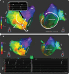
Atypical atrial flutter: potential mechanisms and the effect of reversible PF pulses. A, When the left atrium was mapped during the spontaneous atypical atrial flutter in patient number 1, the activation pattern was consistent with 2 potential mechanisms: (1) a focal tachycardia with a site of origin in the low anterior left atrium near the mitral valve (left) or (2) a macro-reentrant flutter circumventing around the mitral annulus (right). In these activation maps, the color bar indicates the order of activation with the earliest being in red and the latest in purple (see Video S2 for a propagation map that more clearly depicts these potential mechanisms). Note in the inset the highly fractionated, long-duration electrograms observed on the catheter mini-electrodes, which contribute to the confusion on the mechanism since they complicate proper annotation of local timing. The left atrial shell is shown in the anteroposterior (left) and left anterior oblique caudal (right) views. B, A reversible pulsed electric field (PFREV) application at the yellow tag terminated the ongoing rhythm. The views of the left atrium are the same as in A. CS, coronary sinus; LAA, left atrial appendage; LPVs, left pulmonary veins; Map, mapping catheter; RIPV, right inferior pulmonary vein; RSPV, right superior pulmonary vein.
Figure 6.
Figure 6.
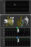
Identification of the slow pathway region to guide ablation of atrioventricular nodal tachycardia. A, In patient number 3, after the atrial fibrillation ablation lesion set, this supraventricular tachycardia (500 ms=120 bpm) occurred spontaneously and was demonstrated to be atrioventricular nodal reentrant tachycardia by standard electrophysiological maneuvers. B, A limited right atrial geometry is shown (transparent yellow) adjacent to the left atrial anatomy (silver) in either right anterior oblique (left) or steep left anterior oblique (right) views. The silver and red tags represent locations where reversible pulsed electric field (PFREV) applications were delivered. This resulted in either (1) termination of the tachycardia, consistent with the site being critical for the tachycardia (red tag; C; Video S6) or (2) no effect on the tachycardia (silver tags; D; Video S7), consistent with not being critical for the tachycardia circuit. CS indicates coronary sinus; HIS, His bundle catheter; LAA, left atrial appendage; LIPV, left inferior pulmonary vein; LSPV, left superior pulmonary vein; Map, mapping catheter; RIPV, right inferior pulmonary vein; and RSPV, right superior pulmonary vein.

Comment in

References

    1. Patel AM, d’Avila A, Neuzil P, Kim SJ, Mela T, Singh JP, Ruskin JN, Reddy VY. Atrial tachycardia after ablation of persistent atrial fibrillation: identification of the critical isthmus with a combination of multielectrode activation mapping and targeted entrainment mapping. Circ Arrhythmia Electrophysiol. 2008;1:14–22. doi: 10.1161/CIRCEP.107.748160 - PubMed
    1. Batista Napotnik T, Polajžer T, Miklavčič D. Cell death due to electroporation - a review. Bioelectrochemistry. 2021;141:107871. doi: 10.1016/j.bioelechem.2021.107871 - PubMed
    1. van Driel VJ, Neven K, van Wessel H, Vink A, Doevendans PA, Wittkampf FH. Low vulnerability of the right phrenic nerve to electroporation ablation. Heart Rhythm. 2015;12:1838–1844. doi: 10.1016/j.hrthm.2015.05.012 - PubMed
    1. Neven K, van Es R, van Driel V, van Wessel H, Fidder H, Vink A, Doevendans P, Wittkampf F. Acute and long-term effects of full-power electroporation ablation directly on the porcine esophagus. Circ Arrhythm Electrophysiol. 2017;10:e004672. doi: 10.1161/CIRCEP.116.004672 - PubMed
    1. Koruth J, Kuroki K, Iwasawa J, Enomoto Y, Viswanathan R, Brose R, Buck ED, Speltz M, Dukkipati SR, Reddy VY. Preclinical evaluation of pulsed field ablation: electrophysiological and histological assessment of thoracic vein isolation. Circ Arrhythm Electrophysiol. 2019;12:e007781. doi: 10.1161/CIRCEP.119.007781 - PMC - PubMed

Publication types