

Intravascular ultrasound to aid in the diagnosis and revision of an intra-aortic pedicle screw: illustrative case
- PMID: 37728279
- PMCID: PMC10555651
- DOI: 10.3171/CASE23272
Intravascular ultrasound to aid in the diagnosis and revision of an intra-aortic pedicle screw: illustrative case
Abstract
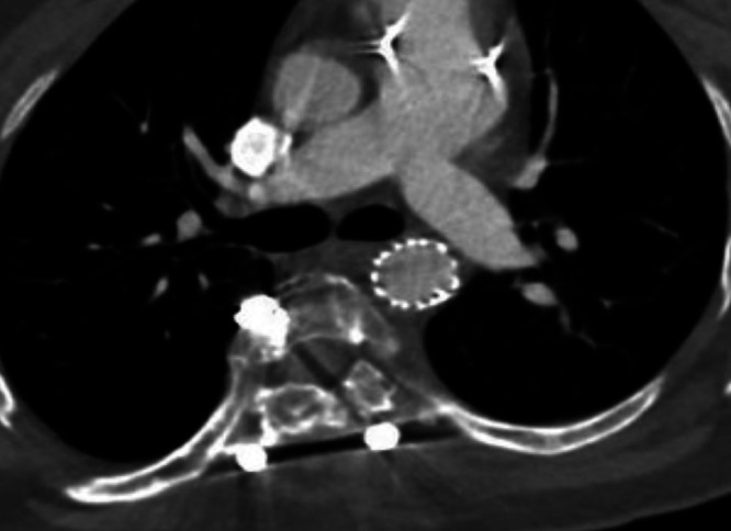
Background: Pedicle screw impingement on vessel walls has the potential for complications due to pulsatile effects and wall erosion. Artifacts from spinal instrumentation create difficulty in accurately evaluating this interface. The authors present the first case of intravascular ultrasound (IVUS) used to characterize a pedicle screw breach into the aortic lumen.
Observations: A 21-year-old female with surgically corrected scoliosis underwent computed tomography angiography (CTA) 3 years postoperatively, which revealed a pedicle screw within the thoracic aorta lumen. Metal artifact distorted the CTA images, which prompted the decision to use intraoperative IVUS. The IVUS confirmed the noninvasive imaging findings and guided final decisions regarding aortic endograft size and location during spine hardware revision.
Lessons: For asymptomatic patients presenting with pedicle screws malpositioned in or near the aorta, treatment decisions revolve around the extent of vessel wall penetration. Intraluminal depth can be obscured by artifact on computed tomography or magnetic resonance imaging or inadequately evaluated by a transesophageal echocardiogram. In our intraoperative experience, IVUS confirmed the depth of vessel lumen violation by a single pedicle screw and no wall penetration by two additional screws of concern. This was useful in deciding on thoracic endovascular aortic repair graft size and landing zone and facilitated safe spinal instrumentation removal and revision.
Keywords: aorta; case report; intravascular ultrasound; pedicle screw; revision.
Conflict of interest statement
Figures


Similar articles
-
Malpositioned pedicle screw in spine deformity surgery endangering the aorta: report of two cases, review of literature, and proposed management algorithm.Spine Deform. 2020 Aug;8(4):809-817. doi: 10.1007/s43390-020-00094-5. Epub 2020 Mar 13. Spine Deform. 2020. PMID: 32170660 Review.
-
Management of a periaortic spinal pedicle screw in a patient with adult presentation thoracic scoliosis.J Orthop Case Rep. 2024 Jan;14(1):40-43. doi: 10.13107/jocr.2024.v14.i01.4140. J Orthop Case Rep. 2024. PMID: 38292094 Free PMC article.
-
Aortic Injury by Thoracic Pedicle Screw. When Is Aortic Repair Required? Literature Review and Three New Cases.World Neurosurg. 2019 Aug;128:216-224. doi: 10.1016/j.wneu.2019.04.173. Epub 2019 May 9. World Neurosurg. 2019. PMID: 31077895 Review.
-
Evaluation of pedicle screw placement in the deformed spine using intraoperative plain radiographs: a comparison with computerized tomography.Spine (Phila Pa 1976). 2005 Sep 15;30(18):2084-8. doi: 10.1097/01.brs.0000178818.92105.ec. Spine (Phila Pa 1976). 2005. PMID: 16166900
-
Delayed open treatment of aortic penetration by a thoracic pedicle screw: illustrative case.J Neurosurg Case Lessons. 2023 Feb 27;5(9):CASE22533. doi: 10.3171/CASE22533. Print 2023 Feb 27. J Neurosurg Case Lessons. 2023. PMID: 36852772 Free PMC article.
References
-
- Alomari S, Lubelski D, Lehner K, et al. Safety and accuracy of freehand pedicle screw placement and the role of intraoperative O-arm: a single-institution experience. Spine (Phila Pa 1976) 2023;48(3):180–188. - PubMed
-
- Kayacı S, Cakir T, Dolgun M, et al. Aortic injury by thoracic pedicle screw. When is aortic repair required? Literature review and three new cases. World Neurosurg. 2019;128:216–224. - PubMed
-
- Szolar DH, Preidler KW, Steiner H, et al. Vascular complications in lumbar disk surgery: report of four cases. Neuroradiology. 1996;38(6):521–525. - PubMed
-
- Bingol H, Cingoz F, Yilmaz AT, Yasar M, Tatar H. Vascular complications related to lumbar disc surgery. J Neurosurg. 2004;100(3 Suppl Spine):249–253. - PubMed
LinkOut - more resources
Full Text Sources
