

Single-level ossified ligamentum flavum causing a holocord syrinx: illustrative case
- PMID: 37728291
- PMCID: PMC10555613
- DOI: 10.3171/CASE23340
Single-level ossified ligamentum flavum causing a holocord syrinx: illustrative case
Abstract
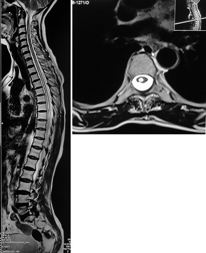
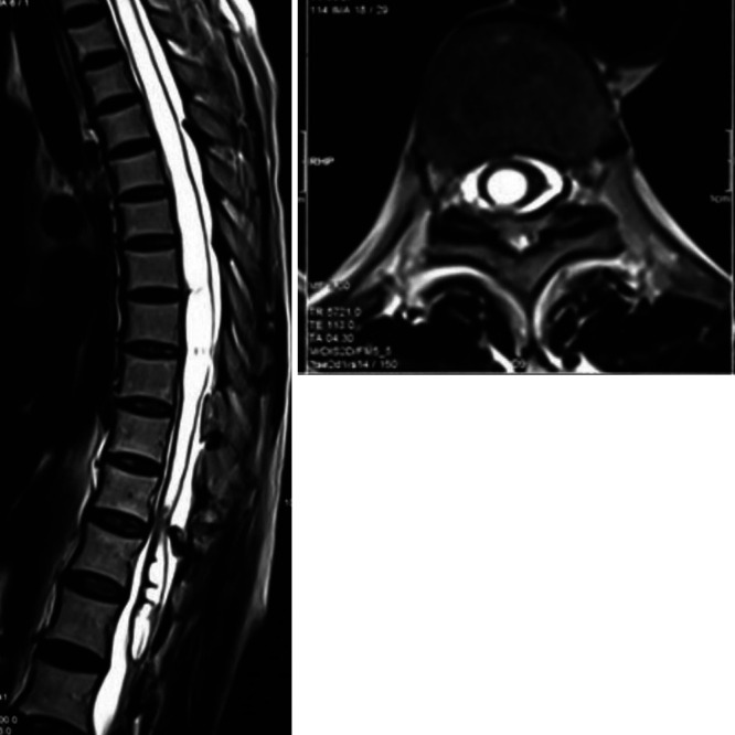
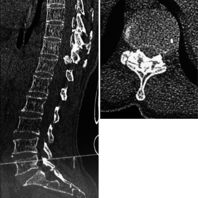
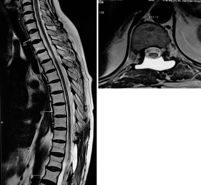
Background: Syringomyelia is a neurological disorder that is caused by abnormal cerebrospinal fluid flow or circulation. It is an incidental finding in most cases, predominantly presenting with sensory symptoms of insensitivity to pain and temperature. Spinal ossified ligamentum flavum (OLF) leading to syringomyelia is one of the rare causes. The authors report an unusual case of syringomyelia due to a thoracic OLF.
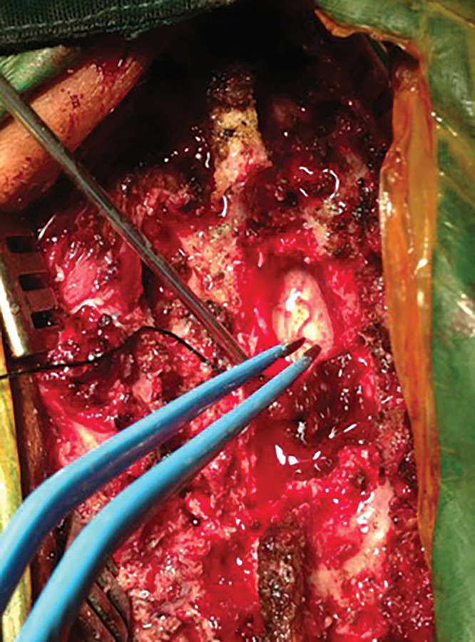
Observations: A 54-year-old female presented with backache, difficulty walking, spasticity in the bilateral lower limbs, tingling sensation in the bilateral lower limbs, and paraparesis for 5 years. Her radiological investigations were suggestive of an OLF causing a syrinx. She underwent laminectomy, and her syrinx resolved on subsequent follow-up.
Lessons: A syrinx due to a single-level OLF is rare, and this uncommon cause should be kept in mind while formulating treatment plans.
Keywords: OLF; ossified ligamentum flavum; syringomyelia; syrinx.
Conflict of interest statement
Figures
References
-
- Greitz D. Unraveling the riddle of syringomyelia. Neurosurg Rev. 2006;29(4):251–264. - PubMed
-
- Milhorat TH, Chou MW, Trinidad EM, et al. Chiari I malformation redefined: clinical and radiographic findings for 364 symptomatic patients. Neurosurgery. 1999;44(5):1005–1017. - PubMed
-
- Roy AK, Slimack NP, Ganju A. Idiopathic syringomyelia: retrospective case series, comprehensive review, and update on management. Neurosurg Focus. 2011;31(6):E15. - PubMed
-
- Klekamp J, Batzdorf U, Samii M, Bothe HW. Treatment of syringomyelia associated with arachnoid scarring caused by arachnoiditis or trauma. J Neurosurg. 1997;86(2):233–240. - PubMed
-
- Roser F, Ebner FH, Sixt C, Hagen JM, Tatagiba MS. Defining the line between hydromyelia and syringomyelia. A differentiation is possible based on electrophysiological and magnetic resonance imaging studies. Acta Neurochir (Wien) 2010;152(2):213–219. discussion 219. - PubMed
LinkOut - more resources
Full Text Sources
