

Extradural lumbar arteriovenous fistula imitating a synovial cyst: illustrative case
- PMID: 37728292
- PMCID: PMC10555559
- DOI: 10.3171/CASE23280
Extradural lumbar arteriovenous fistula imitating a synovial cyst: illustrative case
Abstract
Background: Spinal dural arteriovenous fistula is the most common spinal vascular malformation. It usually presents with neurological deficits due to venous congestive myelopathy. Sometimes, however, the symptoms are radicular and can imitate those of a disc hernia or synovial cyst.
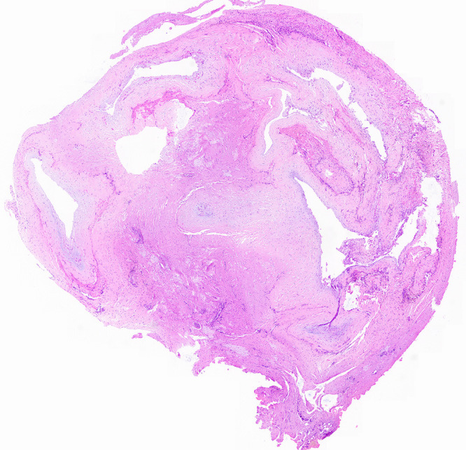
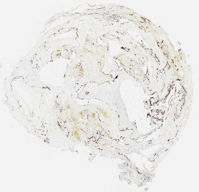
Observations: In this illustrated case, a 34-year-old male patient presented with radicular pain due to nerve root compression. In the magnetic resonance imaging (MRI) examination of the spine, a synovial cyst was suspected, so the patient underwent no further examination before surgery. Intraoperatively, the authors were surprised to see a vascular malformation, which was shown to be an extradural arteriovenous fistula according to the histopathological examination.
Lessons: In atypical MRI findings of the spine, vascular malformations should be considered. In cases with a spinal dural arteriovenous fistula, the thrombosis risk can be high and lead to a need for prolonged anticoagulation.
Keywords: extradural; spinal fistula; synovial cyst.
Conflict of interest statement
Figures

References
-
- Mamaril-Davis J, Aguilar-Salinas P, Avila MJ, Dumont T, Avery MB. Recurrence rates following treatment of spinal vascular malformations: a systematic review and meta-analysis. World Neurosurg. 2023;173:e250–e297. - PubMed
-
- Maimon S, Luckman Y, Strauss I. Spinal dural arteriovenous fistula: a review. Adv Tech Stand Neurosurg. 2016;(43):111–137. - PubMed
-
- Thron A. Spinal dural arteriovenous fistulas. Radiologe. 2001;41(11):955–960. - PubMed
-
- Gioppo A, Faragò G, Giannitto C, et al. Sacral dural arteriovenous fistulas: a diagnostic and therapeutic challenge - single-centre experience of 13 cases and review of the literature. J Neurointerv Surg. 2018;10(4):415–421. - PubMed
-
- Reinges MHT, Thron A, Mull M, Huffmann BC, Gilsbach JM. Dural arteriovenous fistulae at the foramen magnum. J Neurol. 2001;248(3):197–203. - PubMed
LinkOut - more resources
Full Text Sources
