Spontaneous remission of skull Langerhans cell histiocytosis that had developed by repeated head injury: illustrative case
- PMID: 37728298
- PMCID: PMC10555564
- DOI: 10.3171/CASE2327
Spontaneous remission of skull Langerhans cell histiocytosis that had developed by repeated head injury: illustrative case
Abstract
Background: Langerhans cell histiocytosis (LCH) was previously characterized as the proliferation of Langerhans-type histiocytes with a wide range of clinical presentations that arise mostly in children. The typical presentation is a gradually enlarging, painless skull mass. Rapid clinical deterioration is rare.
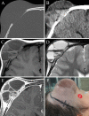
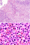
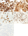
Observations: A 3-year-old boy who had incurred a right frontal impact head injury demonstrated no apparent neurological deficits. He subsequently bruised the same region multiple times. The right frontal swelling gradually increased over the course of 6 days after the initial injury. Skull radiography showed no bony lesion. The same site enlarged markedly 12 days after the initial injury. Magnetic resonance imaging revealed a frontal bony tumorous lesion associated with multiple subcutaneous cystic mass lesions. The patient underwent open biopsy of the skull lesion and evacuation of the subcutaneous lesions. Histopathological examination confirmed the diagnosis of LCH. Immunohistochemical evaluation revealed positivity for CD1a and langerin and no immunopositivity for BRAF V600E. The skull lesion spontaneously disappeared 30 days after the biopsy without recurrence.
Lessons: Physicians should be aware of this rare clinical manifestation of LCH that developed by a repeat head injury.
Keywords: head injury; inflammatory cytokine; local inflammatory response; skull Langerhans cell histiocytosis; spontaneous remission.
Conflict of interest statement
Figures


Similar articles
-
Spontaneous extradural hemorrhage due to Langerhans cell histiocytosis of the skull in a child: A rare presentation.J Pediatr Neurosci. 2016 Jan-Mar;11(1):52-5. doi: 10.4103/1817-1745.181248. J Pediatr Neurosci. 2016. PMID: 27195034 Free PMC article.
-
Coexistence of intracranial Langerhans cell histiocytosis and Erdheim-Chester disease in a pediatric patient: a case report.Childs Nerv Syst. 2016 May;32(5):893-6. doi: 10.1007/s00381-015-2929-6. Epub 2015 Oct 14. Childs Nerv Syst. 2016. PMID: 26466952
-
Aggressive unifocal bone Langerhans cell histiocytosis with soft tissue extension both responsive to radiotherapy: a case report.Radiat Oncol. 2022 Aug 1;17(1):137. doi: 10.1186/s13014-022-02108-0. Radiat Oncol. 2022. PMID: 35915468 Free PMC article.
-
Langerhans cell histiocytosis with aneurysmal bone cyst-like changes: a case-based literature review.Childs Nerv Syst. 2023 Nov;39(11):3057-3064. doi: 10.1007/s00381-023-06108-7. Epub 2023 Jul 31. Childs Nerv Syst. 2023. PMID: 37522932 Free PMC article. Review.
-
A common presentation - turning out as an uncommon diagnosis: From hip pain to Langerhans cell histiocytosis.Am J Med Sci. 2022 Sep;364(3):353-358. doi: 10.1016/j.amjms.2022.04.014. Epub 2022 Apr 25. Am J Med Sci. 2022. PMID: 35472335 Review.
Cited by
-
Pediatric Eosinophilic Granuloma Associated With Delayed Epidural Hematoma Following on Seizure: A Case Report.Brain Tumor Res Treat. 2024 Apr;12(2):148-151. doi: 10.14791/btrt.2024.0018. Brain Tumor Res Treat. 2024. PMID: 38742265 Free PMC article.
References
-
- Lee KW, McLeary MS, Zuppan CW, Won DJ. Langerhans’ cell histiocytosis presenting with an intracranial epidural hematoma. Pediatr Radiol. 2000;30(5):326–328. - PubMed
-
- Mosiewicz A, Rola R, Jarosz B, Trojanowska A, Trojanowski T. Langerhans cell histiocytosis of the parietal bone with epidural and extracranial expansion: case report and a review of the literature. Neurol Neurochir Pol. 2010;44(2):196–203. - PubMed
-
- Martínez-Lage JF, Bermúdez M, Martínez-Barba E, Fuster JL, Poza M. Epidural hematoma from a cranial eosinophilic granuloma. Childs Nerv Syst. 2002;18(1–2):74–76. - PubMed
LinkOut - more resources
Full Text Sources
Research Materials

