

A new strategy for treating drug-resistant focal aware seizures: thalamic specific nuclei deep brain stimulation. Illustrative case
- PMID: 37728299
- PMCID: PMC10555561
- DOI: 10.3171/CASE23303
A new strategy for treating drug-resistant focal aware seizures: thalamic specific nuclei deep brain stimulation. Illustrative case
Abstract
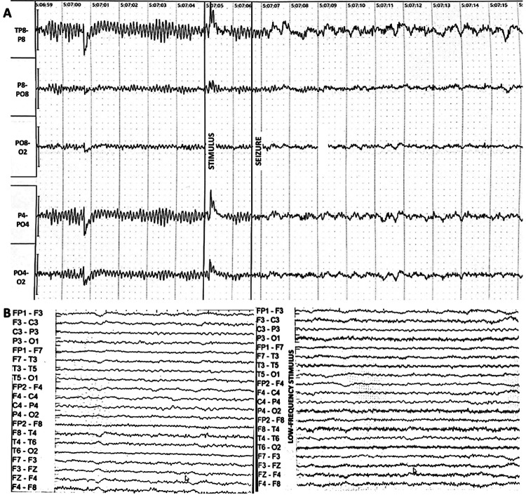
Background: Focal aware seizures (FASs) are relatively common and frequently pharmaco-resistant. If the seizure onset zone (SOZ) is in eloquent cortical areas, making resective surgery risky and inadvisable, deep brain stimulation (DBS) of the anterior nucleus of the thalamus, which is efficacious in less than half of the cases, has been the main alternative. New targets should be searched to address this deficiency. The present study aims to determine if DBS of different thalamic specific nuclei can modulate the abnormal electrical activity of the SOZ located in their respective cortical projection areas. Herein, the authors present the first patient in an ongoing trial.
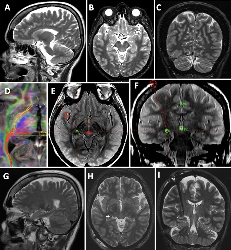
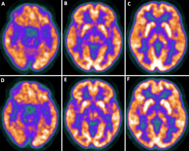
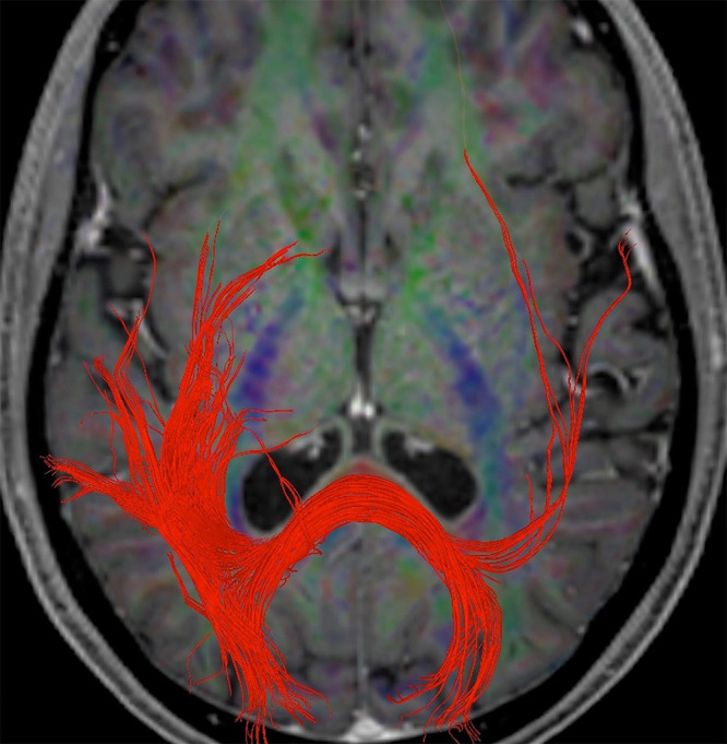
Observations: A 60-year-old female patient presented with 25-year history of pharmaco-resistant focal aware visual seizures frequently evolving to focal impaired awareness seizures. The SOZ was in the right occipital lobe (positron emission tomography-computed tomography/video electroencephalography). Magnetic resonance imaging was normal. She underwent ipsilateral lateral geniculate nucleus (LGN) DBS procedure. After a 24-month follow-up, seizure frequency decreased by 97%, improving quality of life and daily functioning without complications.
Lessons: This is the first time the LGN has been targeted in humans. The results support the hypothesis that led to this study. This strategy represents a paradigm shift in the way of treating pharmaco-resistant FASs not amenable to resective surgery.
Keywords: deep brain stimulation; epilepsy; focal aware seizures; lateral geniculate nucleus; thalamic specific nuclei.
Conflict of interest statement
Figures
Similar articles
-
Ventral intermediate nucleus deep brain stimulation for treatment-resistant focal aware motor seizures: illustrative case.J Neurosurg Case Lessons. 2023 Apr 3;5(14):CASE2320. doi: 10.3171/CASE2320. Print 2023 Apr 3. J Neurosurg Case Lessons. 2023. PMID: 37014006 Free PMC article.
-
Deep brain stimulation of thalamus for epilepsy.Neurobiol Dis. 2023 Apr;179:106045. doi: 10.1016/j.nbd.2023.106045. Epub 2023 Feb 20. Neurobiol Dis. 2023. PMID: 36809846 Review.
-
Clinical efficacy and safety of anterior thalamic deep brain stimulation for intractable drug resistant epilepsy.Epilepsy Res. 2023 Sep;195:107199. doi: 10.1016/j.eplepsyres.2023.107199. Epub 2023 Jul 27. Epilepsy Res. 2023. PMID: 37531721
-
Deep brain stimulation in patients with long history of drug resistant epilepsy and poor functional status: Outcomes based on the different targets.Clin Neurol Neurosurg. 2021 Sep;208:106827. doi: 10.1016/j.clineuro.2021.106827. Epub 2021 Jul 21. Clin Neurol Neurosurg. 2021. PMID: 34329812
-
Deep brain stimulation for seizure control in drug-resistant epilepsy.Neurosurg Focus. 2018 Aug;45(2):E4. doi: 10.3171/2018.4.FOCUS1872. Neurosurg Focus. 2018. PMID: 30064326 Review.
References
-
- World Health Organization. Epilepsy. [internet]. Geneva, Switzerland. [update 9 February 2022]. Accessed November 17, 2022. https://www.who.int/news-room/fact-sheets/detail/epilepsy#.
-
- Beghi E. The epidemiology of epilepsy. Neuroepidemiology. 2020;54(2):185–191. - PubMed
-
- Hauser WA, Annegers JF, Rocca WA. Descriptive epidemiology of epilepsy: contributions of population-based studies from Rochester, Minnesota. Mayo Clin Proc. 1996;71(6):576–586. - PubMed
-
- Krauss GL, Klein P, Brandt C, et al. Safety and efficacy of adjunctive cenobamate (YKP3089) in patients with uncontrolled focal seizures: a multicentre, double-blind, randomised, placebo-controlled, dose-response trial. Lancet Neurol. 2020;19(1):38–48. - PubMed
-
- Li S, Ding D, Wu J. Definitions and epidemiology of epilepsy. In: Shorvon S, Guerrini R, Cook M, Lhatoo SD, editors. Oxford Textbook of Epilepsy and Epileptic Seizure. Oxford University Press; 2013. pp. 51–60.
LinkOut - more resources
Full Text Sources
