. 2023 Oct 11;137(19):1513-1531.
doi: 10.1042/CS20230804.

Myeloid cell-specific deletion of epidermal growth factor receptor aggravates acute cardiac injury

Affiliations

Myeloid cell-specific deletion of epidermal growth factor receptor aggravates acute cardiac injury

Ama D Okyere et al. Clin Sci (Lond). .

Abstract

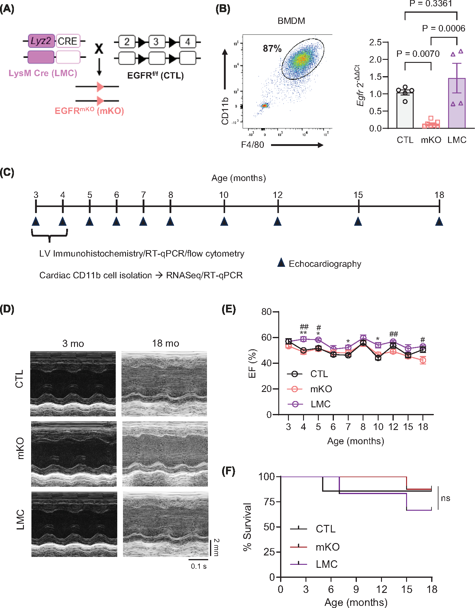
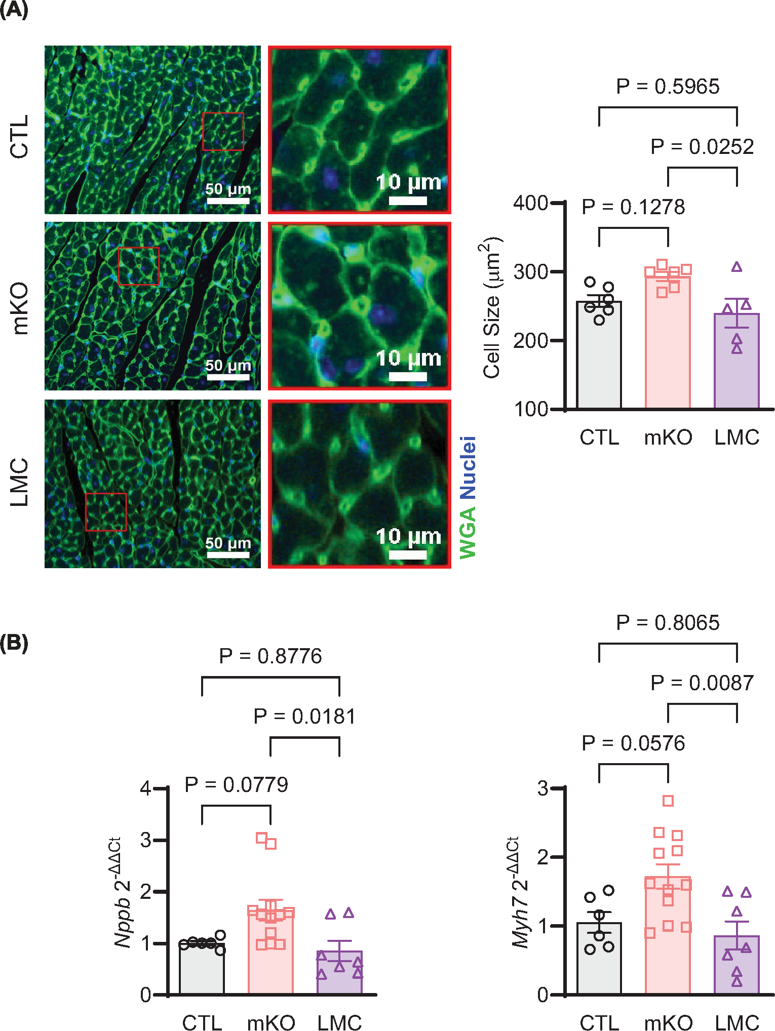
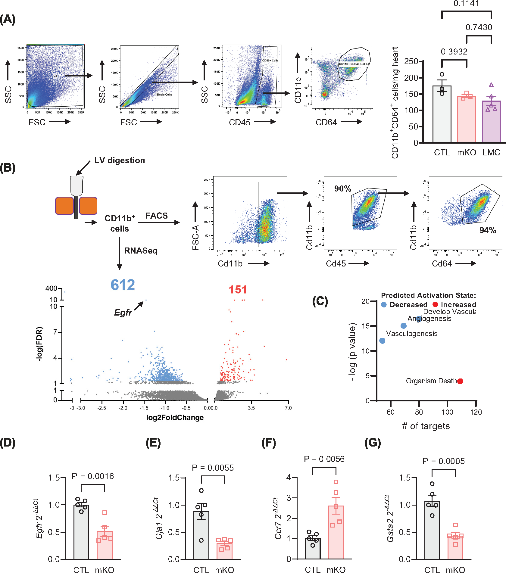
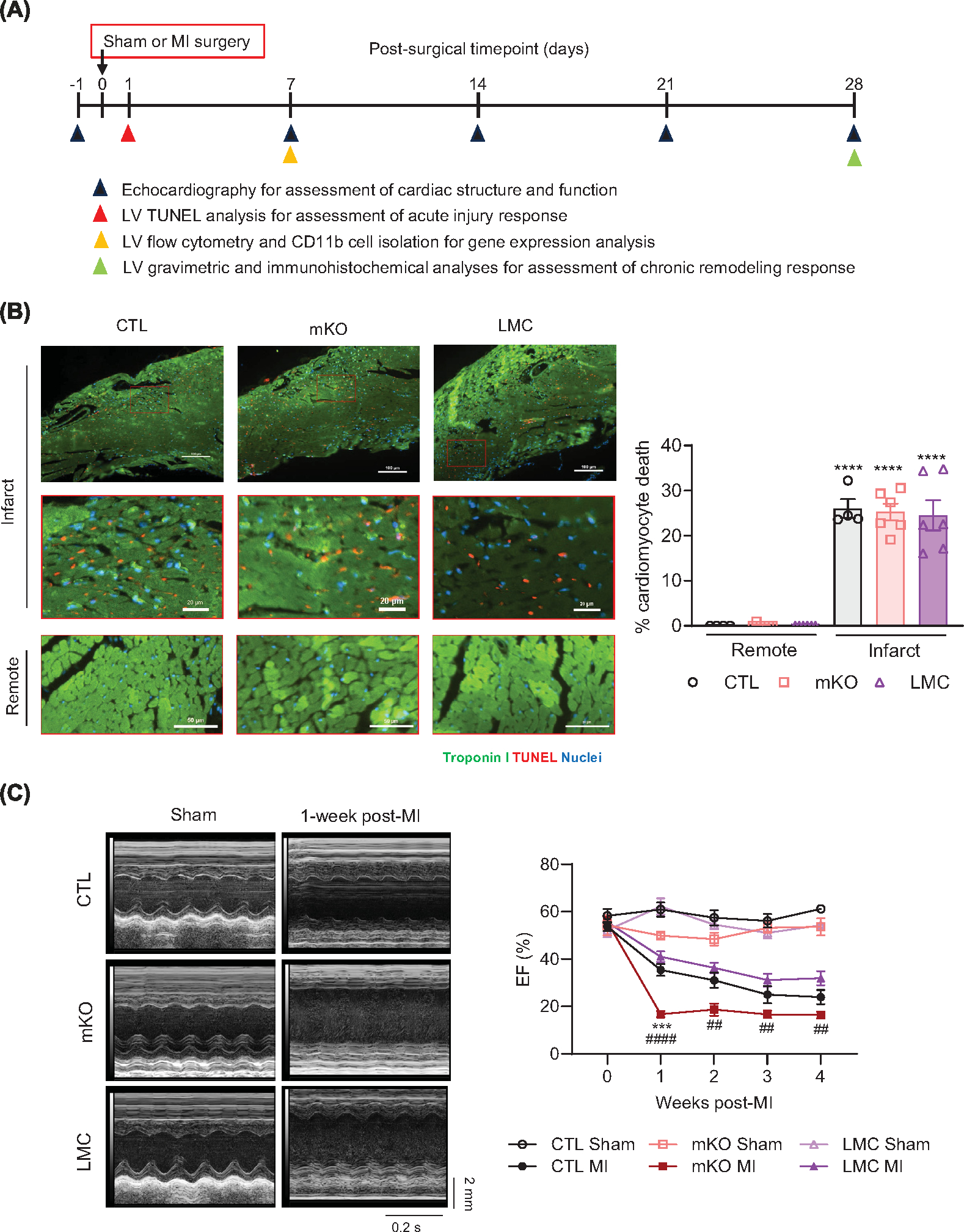
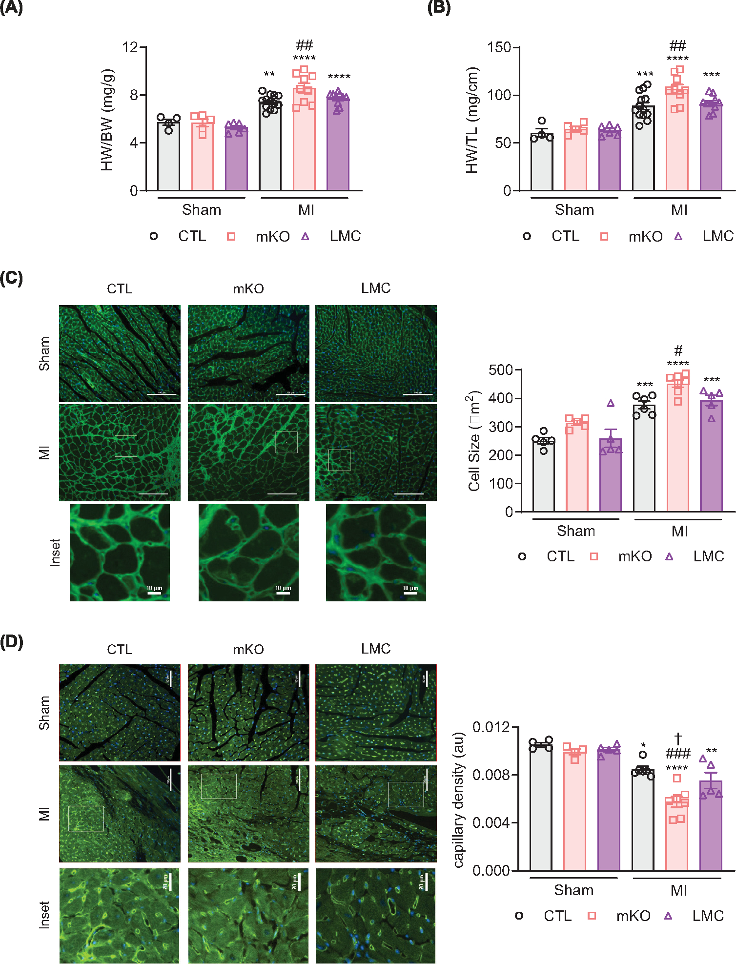
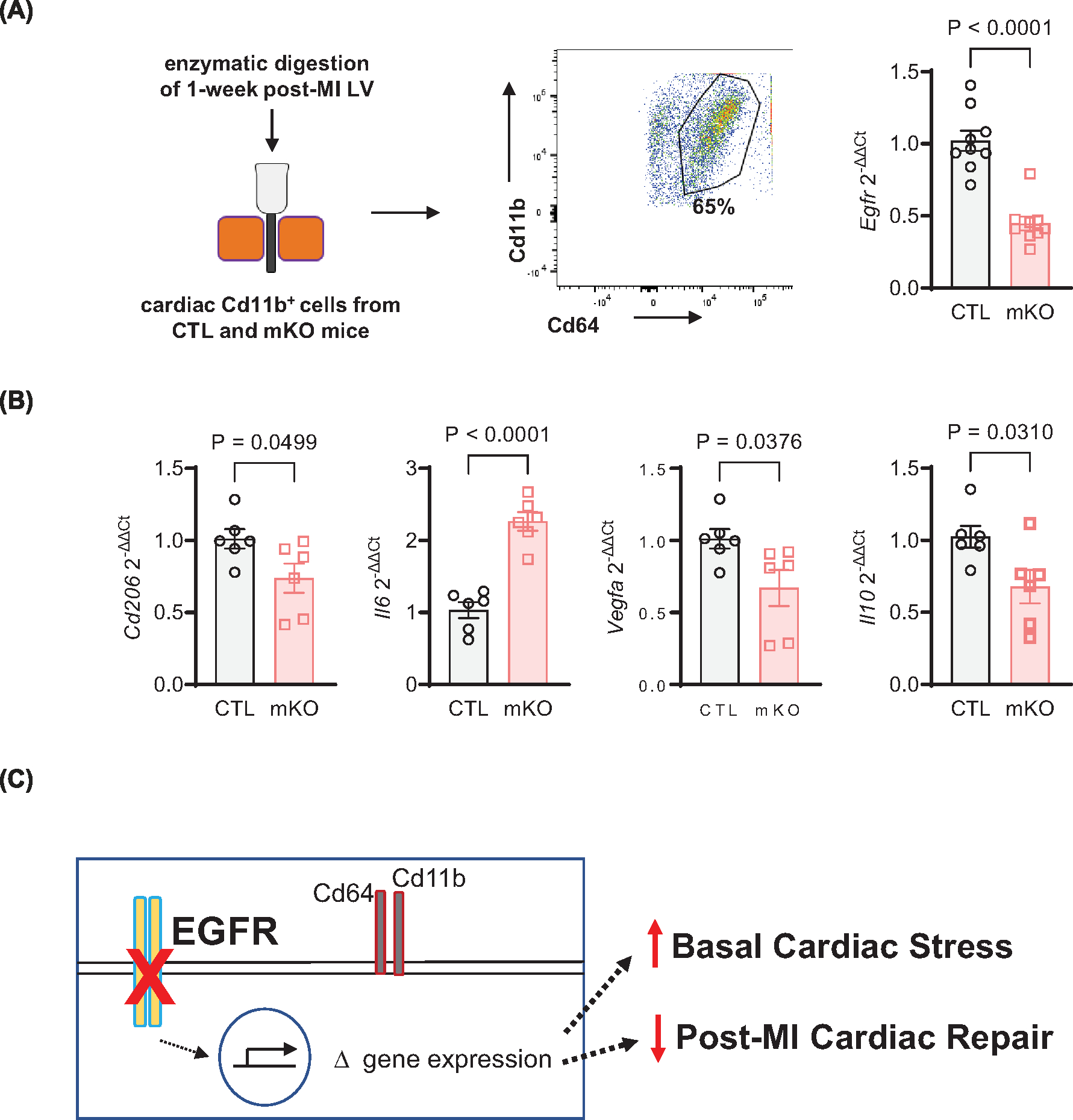
Myeloid cells, including macrophages, play important roles as first responders to cardiac injury and stress. Epidermal growth factor receptor (EGFR) has been identified as a mediator of macrophage responsiveness to select diseases, though its impact on cardiac function or remodeling following acute ischemic injury is unknown. We aimed to define the role of myeloid cell-specific EGFR in the regulation of cardiac function and remodeling following acute myocardial infarction (MI)-induced injury. Floxed EGFR mice were bred with homozygous LysM-Cre (LMC) transgenic mice to yield myeloid-specific EGFR knockout (mKO) mice. Via echocardiography, immunohistochemistry, RNA sequencing and flow cytometry, the impact of myeloid cell-specific EGFR deletion on cardiac structure and function was assessed at baseline and following injury. Compared with LMC controls, myeloid cell-specific EGFR deletion led to an increase in cardiomyocyte hypertrophy at baseline. Bulk RNASeq analysis of isolated cardiac Cd11b+ myeloid cells revealed substantial changes in mKO cell transcripts at baseline, particularly in relation to predicted decreases in neovascularization. In response to myocardial infarction, mKO mice experienced a hastened decline in cardiac function with isolated cardiac Cd11b+ myeloid cells expressing decreased levels of the pro-reparative mediators Vegfa and Il10, which coincided with enhanced cardiac hypertrophy and decreased capillary density. Overall, loss of EGFR qualitatively alters cardiac resident macrophages that promotes a low level of basal stress and a more rapid decrease in cardiac function along with worsened repair following acute ischemic injury.

Keywords: epidermal growth factor receptor; macrophages; myeloid cell; myocardial remodeling.


Figures

Figure 1.
Figure 1.. Myeloid cell-specific EGFR deletion does not negatively impact cardiac function over time.
(A) Schematic showing generation of myeloid cell-specific EGFR knockout mice (EGFRmKO, mKO). Homozygous LysM-Cre transgenic mice (LMC) were crossed to mice carrying a conditional allele of Egfr with exon 3 flanked by two loxP sites (EGFRf/f, control (CTL)). (B) Bone marrow derived macrophages (BMDM) were subject to flow cytometry to verify macrophage (mf) derivation using CD11b, and F4/80. RT-qPCR was used to measure Egfr in mKO versus CTL and LMC BMDM, normalized to Gapdh. N = 5 (CTL), 7 (mKO) or 4 (LMC); data are mean ± SEM, one-way ANOVA with Tukey’s multiple comparisons test. (C) Timeline of comparative assessments of CTL, mKO and LMC mice via echocardiography, immunohistochemistry, RNASeq and RT-qPCR analyses. (D) Representative m-mode echocardiograms of CTL, mKO and LMC hearts at 3 and 18 mo. Upper scale depicts 2 mm. Bottom scale depicts 0.1 seconds. (E) Ejection fraction (EF, %) of CTL, mKO and LMC mouse hearts measured via echocardiography from 3 to 18 months of age. Data are mean ± SEM; *P<0.05 CTL vs. LMC, #P<0.05, ##P<0.01, two-way repeated measures ANOVA to determine the row (timepoint) × column (genotype/age) factor interaction (P=0.1445) with Tukey’s multiple comparisons post-hoc analyses at the individual timepoints. N = 8 (CTL), 7 (mKO) or 6 (LMC). (F) Survival analysis of CTM, mKO and LMC mice until 18 months of age; ns = not significant, Log-rank (Mantel-Cox) test. Initial N = 8 (CTL), 7 (mKO) and 6 (LMC).
Figure 2.
Figure 2.. Myeloid cell-specific EGFR deletion induces mild cardiomyocyte hypertrophy.
(A) Cardiomyocyte size was assessed in the LV of 12- to 16-week-old CTL, mKO and LMC mice after WGA staining. Panels shown are representative regions of interest (ROI) from 20× magnification of the LV from each group, scale bar denotes 50 (left) or 10 (right) μm. Quantification of cardiomyocyte size is shown in the histogram. N = 6 (CTL, mKO) or 5 (LMC); data are mean ± SEM, one-way ANOVA with Tukey’s multiple comparisons test. (B) RT-qPCR was used to measure the expression of Nppb (left) and Myh7 (right) in the LV of 12- to 16-week-old CTL, mKO or LMC mice, normalized to Gapdh. N = 6 (CTL), 12 (mKO) or 7 (LMC); data are mean ± SEM, one-way ANOVA with Tukey’s multiple comparisons test.
Figure 3.
Figure 3.. Myeloid cell-specific EGFR deletion alters the cardiac myeloid cell transcriptome.
(A) Cardiac non-myocytes isolated from 12-week-old CTL, mKO or LMC hearts at baseline underwent flow cytometry analysis for CD11b+CD64+cardiac mf, as quantified in the accompanying histogram. N = 3 (CTL, mKO) and 5 (LMC); data are mean +− SEM, one-way ANOVA with Tukey’s multiple comparisons test. (B) CD11b+ cells were isolated from CTL or mKO hearts via magnetic bead separation, of which >90% were confirmed to be mf as shown via FACS, and then subjected to bulk RNAseq analyses. Isolated Cd11b+ cells from 3 to 6 CTL or mKO mouse hearts per replicate were combined. Volcano plot of differentially expressed RNA transcripts highlighting the number of genes increased and decreased in expression out of total transcripts analyzed. Transcripts were only considered significantly differentially expressed at cutoffs: fold change absolute value (>1.5) and false discovery rate (<0.05). Significant transcripts are coded in blue (down-regulated) or red (up-regulated). (C) IPA analysis grouped transcripts select decreased and increased biological functions predicted by IPA analysis that are altered in mKO versus CTL cardiac myeloid cells. The y axis indicates level of significance, and the × axis indicates number of targets assigned to biological functions. Blue dots indicate decreased activation, and red dot indicates increased activation. RT-qPCR was used to measure Egfr (D), Gja1 (E), Ccr7 (F) and Gata2 (G) expression in CTL and mKO cardiac myeloid cells, normalized to Gapdh. N = 5 (CTL, mKO); data are mean ± SEM, unpaired t-test.
Figure 4.
Figure 4.. Myeloid cell-specific EGFR deletion hastens cardiac dysfunction following myocardial infarction.
(A) Timeline of comparative assessments of CTL, mKO and LMC mice following sham or MI surgery via echocardiography, immunohistochemistry, flow cytometry and RT-qPCR analyses. (B) Representative panels and insets of TUNEL (red)-, and troponin I (Green) and NucBlue (blue)-stained LVs at 24 post-MI in CTL, mKO and LMC hearts. Scale bar denotes 100 μm (infarct), 20 μm (infarct insets) and 50 μm (remote). Accompanying histogram details quantification of cardiomyocyte death at 24 h post-MI in the infarcts versus remote zones. N = 4 (CTL) or 6 (mKO, LMC); data are mean ± SEM; ****P<0.0001 versus remote zone of same genotype, one-way ANOVA with Tukey’s multiple comparisons test. (C) Representative m-mode echocardiograms of CTL, mKO and LMC hearts 1 week following sham or MI surgery. Upper scale depicts 2 mm. Bottom scale depicts 0.2 s. Histogram depicts ejection Fraction (EF, %) of CTL, mKO and LMC mouse hearts prior to (week 0), and 1–4 weeks post-MI. Two-way repeated measures ANOVA was used to determine the row (timepoint) × column (genotype/surgical condition) factor interaction P<0.001. Tukey’s multiple comparisons test was subsequently used to determine statistical significance between genotype/surgical conditions at the individual timepoints; ns, not significant, ***P<0.001 mKO MI versus CTL MI, ##P<0.01, ####P<0.0001 mKO versus LMC. N = 5 (CTL Sham, mKO Sham), 6 (LMC Sham), 8 (CTL MI, LMC MI) or 7 (mKO MI).
Figure 5.
Figure 5.. Myeloid cell-specific EGFR deletion promotes cardiomyocyte hypertrophy and reduces capillary density following myocardial infarction.
Heart weights from CTL, mKO and LMC mice were normalized to body weight (A) or tibia lengths (B) 4 weeks post-MI. Data are mean ± SEM; **P<0.01, ***P<0.001, ****P<0.0001 versus sham of same genotype, ##P<0.01 versus CTL MI, one-way ANOVA with Tukey’s multiple comparisons test. N = 4 (CTL sham), 5 (mKO sham), 6 (LMC sham), 13 (CTL MI), 9 (mKO MI) or 10 (LMC MI). (C) Representative 20× panels and insets (scale bar: 100 μm, upper panels, and 10 μm, lower insets) of WGA (green) and NucBlue (blue)-stained hearts 4 weeks post-surgery in CTL, mKO and LMC hearts. Histogram shows quantification of cardiomyocyte cell size. Data are mean ± SEM; ***P<0.001, ****P<0.0001 versus sham of same genotype, #P<0.05 versus CTL MI, one-way ANOVA with Tukey’s multiple comparisons test. N = 5 (all shams), 6 (CTL), 7 (mKO) or 5 (LMC). (D) Representative 20× images and insets (scale bar: 100 μm, upper panels, and 20 μm insets) of isolectin B4 (ILB4, green)- and NucBlue (blue)-stained hearts 4 weeks post-surgery in CTL, mKO or LMC hearts. Histogram shows quantification of ILB4-stained capillary density. Data are mean ± SEM; *P<0.05, **P<0.01, ****P<0.0001 versus sham of same genotype, ###P<0.001 versus CTL MI, †P<0.05 versus LMC MI, one-way ANOVA with Tukey’s multiple comparisons test. N = 4 (CTL sham, mKO sham), 5 (LMC sham, LMC MI), 6 (CTL MI) and 7 (mKO MI).
Figure 6.
Figure 6.. EGFR-deficient cardiac myeloid cells have reduced expression of reparative transcripts following MI.
(A) CD11b magnetic beads were applied to cardiac non-myocytes to separate CD11b+ myeloid cells at 7d post-MI using a magnetic column, then subjected to flow cytometry to gate for CD45, CD11b, and CD64 mf. RT-qPCR was used to measure Egfr expression in cardiac CD11b+ myeloid cells from CTL and mKO mice at 7d post-MI, normalized to Gapdh. N = 9 (CTL, mKO); data are mean ± SEM, unpaired t-test. (B) RT-qPCR was used to measure Cd206, Il6, Vegfa and Il10, with Gapdh for normalization, in cardiac CD11b+ cells from CTL and mKO mice at 7d post-MI. N = 6 (CTL, mKO); data are mean ± SEM, unpaired t-test. (C) Myeloid cell-specific deletion of EGFR leads to altered regulation of mf gene expression that contributes to mild basal cardiac stress and a decrease in reparative genes following MI-induced injury, ultimately contributing to an early decrease in cardiac function and delaying neovascularization.

Similar articles

Cited by

References

    1. Alvarez-Argote S and O’Meara CC (2021) The evolving roles of cardiac macrophages in homeostasis, regeneration, and repair. Int. J. Mol. Sci. 22 (15), 7923, 10.3390/ijms22157923 - DOI - PMC - PubMed
    1. Dick SA, Macklin JA, Nejat S, Momen A, Clemente-Casares X, Althagafi MG et al. (2019) Self-renewing resident cardiac macrophages limit adverse remodeling following myocardial infarction. Nat. Immunol. 20, 29–39, 10.1038/s41590-018-0272-2 - DOI - PMC - PubMed
    1. Lavine KJ, Pinto AR, Epelman S, Kopecky BJ, Clemente-Casares X, Godwin J et al. (2018) The macrophage in cardiac homeostasis and disease: JACC Macrophage in CVD Series (Part 4). J. Am. Coll. Cardiol. 72, 2213–2230, 10.1016/j.jacc.2018.08.2149 - DOI - PMC - PubMed
    1. Schulz C, Gomez Perdiguero E, Chorro L, Szabo-Rogers H, Cagnard N, Kierdorf K et al. (2012) A lineage of myeloid cells independent of Myb and hematopoietic stem cells. Science 336, 86–90, 10.1126/science.1219179 - DOI - PubMed
    1. Swirski FK and Nahrendorf M (2018) Cardioimmunology: the immune system in cardiac homeostasis and disease. Nat. Rev. Immunol. 18, 733–744, 10.1038/s41577-018-0065-8 - DOI - PubMed

Publication types