

Management of a challenging dura-embedded anterior inferior cerebellar artery loop during a retrosigmoid hearing-preserving vestibular schwannoma resection: microsurgical technique and operative video. Illustrative case
- PMID: 37728311
- PMCID: PMC10555565
- DOI: 10.3171/CASE23304
Management of a challenging dura-embedded anterior inferior cerebellar artery loop during a retrosigmoid hearing-preserving vestibular schwannoma resection: microsurgical technique and operative video. Illustrative case
Abstract
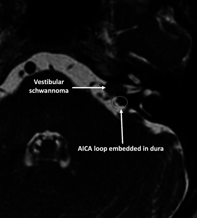
Background: Anatomical variants of the anterior inferior cerebellar artery (AICA), such as an anomalous "AICA loop" embedded in the dura and bone of the subarcuate fossa, increase the complexity and risk of vestibular schwannoma resections. Classically, osseous penetrating AICA loops are the most challenging to mobilize, as the dura must be dissected and the surrounding petrous bone must be drilled to mobilize the AICA away from the surgical corridor and out of harm.
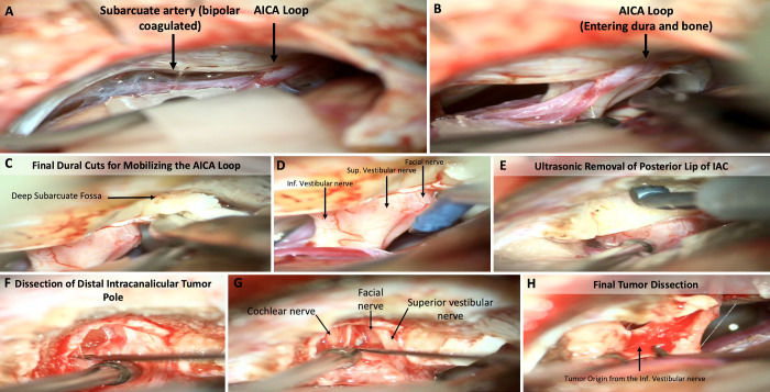
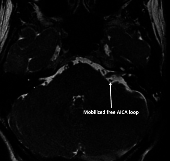
Observations: The authors present a rare case of a dura-embedded, osseous-penetrating AICA loop encountered during a hearing-preserving retrosigmoid approach in which they demonstrate safe and efficient microdissection and mobilization of the AICA loop without having to drill the surrounding bone.
Lessons: Although preoperative recognition of potentially dangerous AICA loops has been challenging, thin-sliced petrous bone computed tomography scanning and high-quality magnetic resonance imaging can be useful in preoperative diagnosis. Furthermore, this report suggests that a retrosigmoid approach is superior, as it allows early intradural recognition and proximal vascular control and facilitates more versatile mobilization of AICA loops.
Keywords: anatomy; anterior inferior cerebellar artery; subarcuate fossa; vestibular schwannoma.
Conflict of interest statement
Figures
Similar articles
-
The anteroinferior cerebellar artery embedded in the subarcuate fossa: a rare anomaly and its clinical significance.Neurosurgery. 2005 Aug;57(2):314-9; discussion 314-9. doi: 10.1227/01.neu.0000166677.70797.5e. Neurosurgery. 2005. PMID: 16094161
-
Challenging Anterior Inferior Cerebellar Artery in Retrosigmoid Vestibular Schwannoma Removal.World Neurosurg. 2019 Jan;121:e370-e378. doi: 10.1016/j.wneu.2018.09.111. Epub 2018 Sep 25. World Neurosurg. 2019. PMID: 30261396
-
The "Deep Subarcuate Fossa" sign and three types of anomalous subarcuate loops encountered during vestibular schwannoma removal.Acta Neurochir (Wien). 2022 Sep;164(9):2483-2490. doi: 10.1007/s00701-022-05288-6. Epub 2022 Jul 1. Acta Neurochir (Wien). 2022. PMID: 35776221
-
Management of anterior inferior cerebellar artery aneurysms: an illustrative case and review of literature.Neurosurg Focus. 2009 May;26(5):E6. doi: 10.3171/2009.1.FOCUS0915. Neurosurg Focus. 2009. PMID: 19409007 Review.
-
[Four cases of direct surgery for anterior inferior cerebellar artery aneurysms].No Shinkei Geka. 2007 Jun;35(6):571-8. No Shinkei Geka. 2007. PMID: 17564050 Review. Japanese.
References
-
- Sekhar LN, Schessel DA, Bucur SD, Raso JL, Wright DC. Partial labyrinthectomy petrous apicectomy approach to neoplastic and vascular lesions of the petroclival area. Neurosurgery. 1999;44(3):537–552. - PubMed
-
- Gannon PJ, Eden AR, Laitman JT. The subarcuate fossa and cerebellum of extant primates: comparative study of a skull-brain interface. Am J Phys Anthropol. 1988;77(2):143–164. - PubMed
-
- Martin RG, Grant JL, Peace D, Theiss C, Rhoton AL., Jr Microsurgical relationships of the anterior inferior cerebellar artery and the facial-vestibulocochlear nerve complex. Neurosurgery. 1980;6(5):483–507. - PubMed
-
- Tanriover N, Rhoton AL., Jr The anteroinferior cerebellar artery embedded in the subarcuate fossa: a rare anomaly and its clinical significance. Neurosurgery. 2005;57(2):314–319. - PubMed
LinkOut - more resources
Full Text Sources
