. 2023 Oct 31;14(5):e0151923.
doi: 10.1128/mbio.01519-23. Epub 2023 Sep 20.

VirB, a key transcriptional regulator of Shigella virulence, requires a CTP ligand for its regulatory activities

Affiliations

VirB, a key transcriptional regulator of Shigella virulence, requires a CTP ligand for its regulatory activities

Taylor M Gerson et al. mBio. .

Abstract

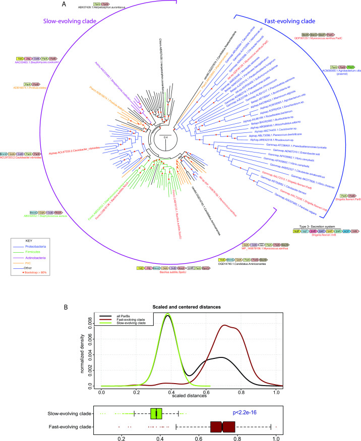
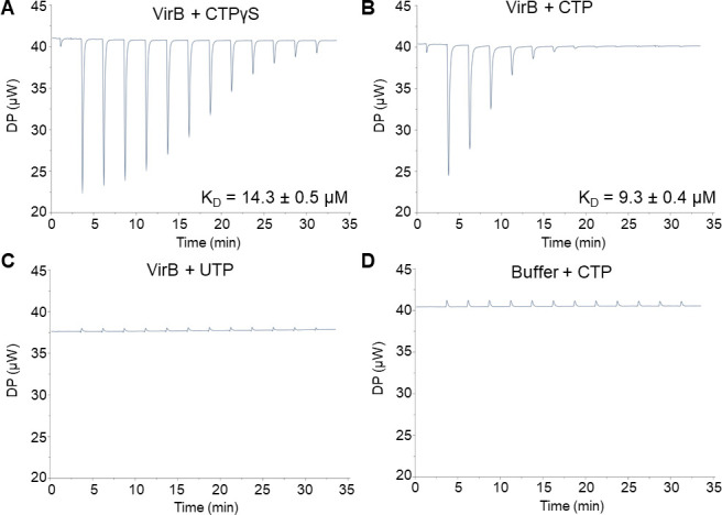
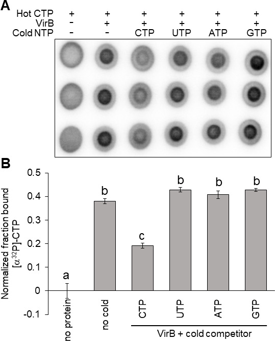
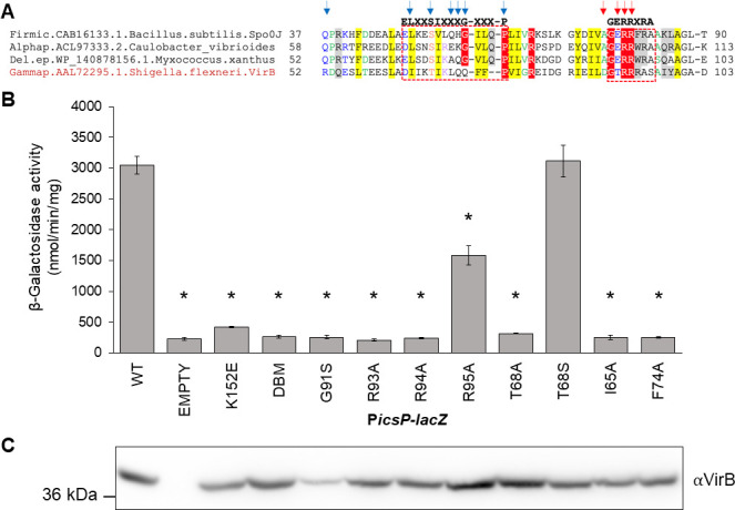
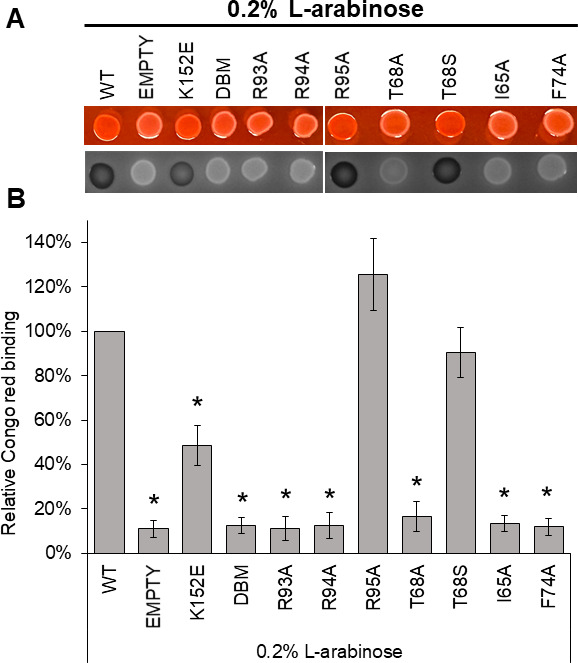
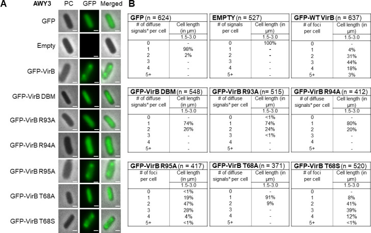
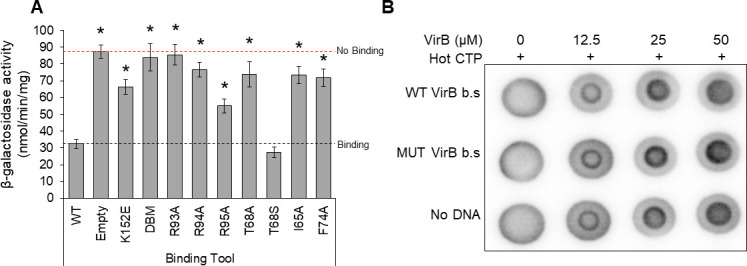
Shigella species cause bacillary dysentery, the second leading cause of diarrheal deaths worldwide. There is a pressing need to identify novel molecular drug targets. Shigella virulence phenotypes are controlled by the transcriptional regulator, VirB. We show that VirB belongs to a fast-evolving, plasmid-borne clade of the ParB superfamily, which has diverged from versions with a distinct cellular role-DNA partitioning. We report that, like classic members of the ParB family, VirB binds a highly unusual ligand, CTP. Mutants predicted to be defective in CTP binding are compromised in a variety of virulence attributes controlled by VirB, likely because these mutants cannot engage DNA. This study (i) reveals that VirB binds CTP, (ii) provides a link between VirB-CTP interactions and Shigella virulence phenotypes, (iii) provides new insight into VirB-CTP-DNA interactions, and (iv) broadens our understanding of the ParB superfamily, a group of bacterial proteins that play critical roles in many bacteria.

Keywords: Congo red phenotype; GFP fusions; ParB/Spo0J; bacterial gene regulation; focus formation; large plasmids; plasmid partitioning; transcriptional silencing/anti-silencing.


Conflict of interest statement

The authors declare no conflict of interest.

Figures

Fig 1
Fig 1
Maximum likelihood tree of ParB protein sequences including the VirB-like fast-evolving sequences. (A) Maximum likelihood tree of ParB protein sequences including the VirB-like fast-evolving sequences. The tree was computed using IQtree and bootstrapped with 1,000 replicates. Nodes and branches corresponding to widely represented phylogenetic clades are colored according to the key. Protein sequences are denoted with their clade, gene, and species name. The slow- and fast-evolving clades are marked and representative gene neighborhoods of distinct clades of the tree are shown. Genes in gene neighborhoods are shown as box arrows with the arrowhead pointing to the gene at the 3′ end. Clade abbreviations include: Actino,: Actinomycetes; Alphap, Alpha-proteobacteria; Betapr, Beta-proteobacteria; candid, Dark-matter bacteria; Chloro, Chloroflexi; del.ep, Delta/epsilon proteobacteria; Firmic, Firmicutes; Gammap, Gamma-proteobacteria; Planct, Planctomycetes. (B) Differential evolutionary rates derived from pairwise centered and scaled maximum-likelihood distances shown as a kernel density and box plots.
Fig 2
Fig 2
The Shigella anti-silencing protein VirB binds a novel ligand, CTP. ITC measurements with (A) 90 µM VirB-His6 and 3 mM CTPγS, (B) 45 µM VirB-His6 and 3 mM CTP, (C) 45 µM VirB-His6 and 3 mM UTP, and (D) VirB binding buffer and 3 mM CTP. In panels B and C, the VirB protein concentration was lowered to 45 µM to reach saturation and conserve protein.
Fig 3
Fig 3
VirB preferentially binds CTP over other NTP ligands found in the cell. (A) DRaCALA images of competition assays assessing the ability of 500 µM indicated cold NTP to compete with binding interactions between 5 nM [α32P]-CTP and 50 µM VirB-His6. Ligand binding is indicated by a darker inner core staining due to rapid immobilization of the VirB-CTP complex. Three independent trials of this experiment were run. Each trial contained three technical replicates. A representative trial is shown (each row represents a technical replicate). (B) Graph of normalized fraction bound (FB) calculated for each sample in Fig. 3A. Significance was calculated using a one-way ANOVA with post hoc Bonferroni, P < 0.05. Lowercase letters indicate statistical groups. Complete statistical analysis provided in Table S3.
Fig 4
Fig 4
Key residues in the predicted CTP binding pocket are required for the anti-silencing activity of VirB. (A) Multiple sequence alignment (MSA of classic ParBs that bind CTP and VirB, generated using the Mafft program with a local pair algorithm. Full alignment and additional details provided in Fig. S2. Taxonomic clade name, Genbank accession, and species name separated by dots are denoted. Consensus abbreviations and coloring scheme are as follows: hydrophobic (FWYILVACM), aromatic (FWY) and aliphatic (ILV) residues shaded yellow; charged (DEHKR) and basic (KRH) residues colored magenta, big (LIFMWYERKQ) residues shaded gray, alcohol-group (ST) residues colored red, polar (STECDRKHNQ) residues colored blue, small (AGSCDNPTV) residues colored green, and tiny (GAS) residues shaded green. Fully conserved residues are shaded red. Arrows indicate nucleotide-binding residues (blue) and catalytic residues (red). (B) β-Galactosidase assay used to assess the regulatory activity of pBAD-virB derivatives at the VirB-dependent icsP promoter. Significance was calculated using a one-way ANOVA with post hoc Tukey HSD, P < 0.05. *, statistically significant compared to wild type. Complete statistical analysis is provided in Table S4. (C) Western Blot analysis using an anti-VirB antibody to assess protein production of pBAD-virB mutants alongside a SeeBlue Plus2 Prestained Standard. Assays were completed with three biological replicates and repeated three times. Representative data are shown.
Fig 5
Fig 5
Key residues in the predicted CTP binding pocket are required for Shigella virulence phenotype. (A) Congo red binding by S. flexneri virB::Tn5 harboring pBAD-virB derivatives under inducing conditions. Images were captured using visible light (top) and blue light (Cy2) (bottom). (B) Quantitative analysis of Congo red binding S. flexneri virB::Tn5 harboring pBAD-virB derivatives (induced). Relative Congo red binding was calculated as [(OD498/OD600)/(average (OD498/OD600)2457T pBAD)] ×100. Assays were completed with three biological replicates and repeated three times. Representative data are shown. Significance was calculated using a one-way ANOVA with post hoc Tukey HSD, P < 0.05. *, statistically significant compared to wild type. Complete statistical analysis is provided in Table S5.
Fig 6
Fig 6
Key residues in the CTP binding pocket are required for VirB focus formation in vivo. (A) Live-cell imaging of GFP-VirB derivatives in a virB mutant strain of S. flexneri (AWY3) induced with 0.02% L-arabinose. Phase-contrast, PC, (left column), fluorescence (middle column) (GFP row, 28 ms exposure; GFP-VirB derivatives and empty rows, 98 ms exposure), and merged (right column). Bars represent 1 µM. (B) Quantification of fluorescent signals observed during live-cell imaging of GFP-VirB derivatives using MicrobeJ. Within tables, a hyphen indicates that no cells were found in this category in any of the images captured; *, maxima detected by MicrobeJ. Complete statistical analysis is provided in Table S6 and field-of-view images are found in Fig. S5.
Fig 7
Fig 7
Characterizing the order of VirB-, CTP-, and DNA-binding site interactions. (A) Ptac promoter activity of pBT-PicsP in the presence of pBAD-virB mutants in a S. flexneri virB::Tn5, as determined by β-galactosidase assays. Assays were completed with three biological replicates and repeated three times. Representative data are shown. Dash lines represent activities generated by the positive and negative controls. Significance was calculated using a one-way ANOVA with post hoc Tukey HSD, *P < 0.05. *, statistically significant compared to wild type. Complete statistical analysis provided in Table S7. Note, K152E and R95A are statistically different from both wild type and the empty plasmid control. (B) DRaCALA DNA dependency assay assessing the ability of VirB to bind CTP in the presence/absence of various DNA substrates. Three technical replicates were completed for each trial and three independent trials of this experiment were run. A representative technical replicate is shown.

Update of

Similar articles

Cited by

References

    1. Hawkins HP. 1909. The identity of British ulcerative colitis and tropical bacillary dysentery. Br Med J 2:1331–1332. doi:10.1136/bmj.2.2549.1331 - DOI - PMC - PubMed
    1. Labrec EH, Schneider H, Magnani TJ, Formal SB. 1964. Epithelial cell penetration as an essential step in the pathogenesis of bacillary dysentery. J Bacteriol 88:1503–1518. doi:10.1128/jb.88.5.1503-1518.1964 - DOI - PMC - PubMed
    1. Niyogi SK. 2007. Increasing antimicrobial resistance--an emerging problem in the treatment of shigellosis. Clin Microbiol Infect 13:1141–1143. doi:10.1111/j.1469-0691.2007.01829.x - DOI - PubMed
    1. Niyogi SK. 2005. Shigellosis. J Microbiol 43:133–143. - PubMed
    1. Weatherspoon-Griffin N, Picker MA, Wing HJ. 2016. The genetic organization and transcriptional regulation of Shigella virulence genes, p 65–107. In Picking WD, Picking WL (ed), Shigella: Molecular and cellular biology. Caister Academic Press, UK. doi:10.21775/9781910190197 - DOI

LinkOut - more resources