. 2023 Sep 20;11(5):e0260423.
doi: 10.1128/spectrum.02604-23. Online ahead of print.

Efficient screening of adsorbed receptors for Salmonella phage LP31 and identification of receptor-binding protein

Affiliations

Efficient screening of adsorbed receptors for Salmonella phage LP31 and identification of receptor-binding protein

Haojie Ge et al. Microbiol Spectr. .

Abstract

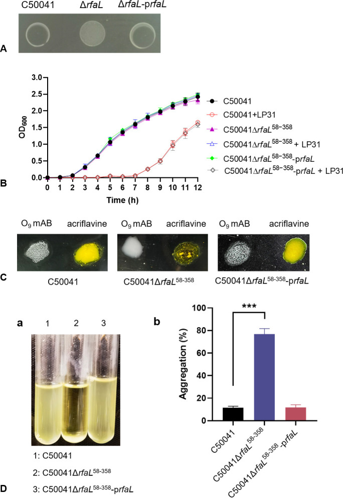
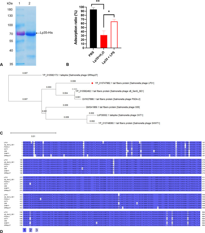
The adsorption process is the first step in the lifecycle of phages and plays a decisive role in the entire infection process. Identifying the adsorption mechanism of phages not only makes phage therapy more precise and efficient but also enables the exploration of other potential applications and modifications of phages. Phage LP31 can lyse multiple Salmonella serotypes, efficiently clearing biofilms formed by Salmonella enterica serovar Enteritidis (S. Enteritidis) and significantly reducing the concentration of S. Enteritidis in chicken feces. Therefore, LP31 has great potential for many practical applications. In this study, we established an efficient screening method for phage infection-related genes and identified a total of 10 genes related to the adsorption process of phage LP31. After the construction of strain C50041ΔrfaL 58-358, it was found that the knockout strain had a rough phenotype as an O-antigen-deficient strain. Adsorption rate and transmission electron microscopy experiments showed that the receptor for phage LP31 was the O9 antigen of S. Enteritidis. Homology comparison and adsorption experiments confirmed that the tail fiber protein Lp35 of phage LP31 participated in the adsorption process as a receptor-binding protein. IMPORTANCE A full understanding of the interaction between phages and their receptors can help with the development of phage-related products. Phages like LP31 with the tail fiber protein Lp35, or a closely related protein, have been reported to effectively recognize and infect multiple Salmonella serotypes. However, the role of these proteins in phage infection has not been previously described. In this study, we established an efficient screening method to detect phage adsorption to host receptors. We found that phage LP31 can utilize its tail fiber protein Lp35 to adsorb to the O9 antigen of S. Enteritidis, initiating the infection process. This study provides a great model system for further studies of how a phage-encoded receptor-binding protein (RBP) interacts with its host's RBP binding target, and this new model offers opportunities for further theoretical and experimental studies to understand the infection mechanism of phages.

Keywords: O9 antigen; Salmonella; adsorption receptor; phage; receptor-binding protein.


Conflict of interest statement

The authors declare no conflict of interest.

Figures

FIG 1
FIG 1
Screening of phage-resistant strains. (A) Comparison before and after killing sensitive bacteria with phage. (B) Screening of the putative phage-resistant mutants by drop spot assay. The red numbers are true phage-resistant mutants. The green numbers are sensitive strains. (C) Growth curves of wild-type C50041 and LP31-resistant mutants with and without phage LP31, cultured in vitro.
FIG 2
FIG 2
Screening of genes related to the phage adsorption process. (A) Analysis of transposon insertion sites in phage LP31-insensitive strains. (B) Schematic structure of LPS, showing that different gene products (color-coded to match panel A) required for LPS synthesis affect the adsorption of phage LP31. Abbreviations are as follows: Glc, glucose; Rha, rhamnose; GlcNAc, N-acetylglucosamine; Man, mannose; Hep, heptose; Gal, galactose; Kdo, 2-keto-3-deoxyoctulosonic acid; P, phosphate; PEtN, phosphoethanolamine. The structure of LPS is that of S. Typhimurium, modified from references (30, 31). (C) Adsorption properties of phage LP31 on S. Enteritidis C50041 or C50041 treated with proteinase K or Ac (NaIO4). (D) Comparative LP31 adsorption measurements on different LP31-resistant mutants. The X-axis is labeled with the various Tn mutants, and they have been color-coded to match the gene identities defined in panel A. **, P < 0.01; ***, P < 0.001.
FIG 3
FIG 3
Characterization of the rfaL gene deletion mutant (C50041ΔrfaL 58-358) and complemented strain (C50041ΔrfaL 58-358-prfaL). (A) The ability of phage LP31 to form clear spots on phage drop spot assays on the wild-type C50041 strain, its rfaL gene deletion mutant variant, and the complemented strain. (B) Growth curves of the wild-type, rfaL deletion mutant, and the complemented mutant cultured in vitro with or without phage LP31. (C) Agglutination phenotypes of the wild-type, rfaL deletion mutant, and complemented mutant. S. Enteritidis with O-antigen supports agglutination by an O9 mAB (monoclonal antibody against O-antigens) but does not agglutinate with acriflavine. The strain with the O-antigen synthesis defect does not agglutinate with O9 mAB but does agglutinate in the presence of acriflavine. (D) The aggregation of different strains: (a) aggregation of bacteria during cultivation; (b) percentage of aggregation of different strains (***, P < 0.001).
FIG 4
FIG 4
Characterization of phage LP31 adsorption. (A) The adsorption of phage LP31 on different wild-type (with O9 antigen) and LPS mutant (without O9 antigen) strains of S. Enteritidis (C50041) and S. Typhimurium (D6). (B) Effect of added S. Enteritidis LPS on the ability of phage LP31 to adsorb to S. Enteritidis C50041. LP31 was added to S. Enteritidis (C50041) with either water or the same volume of LPS (100 mg/mL). (C) Transmission electron microscopy of S. Enteritidis C50041 shows adsorbed phage LP31 (the red arrows are pointing at the phages). 1: Adsorption of phage LP31 to C50041. 2: Lack of adsorption of phage LP31 to C50041ΔrfaL 58−358. 3: Adsorption of phage LP31 to C50041 after incubation with H2O. 4: Lack/decrease of adsorption of phage LP31 to C50041 after incubation with LPS. 5: Lack/decrease of adsorption of phage LP31 to C50041 after incubation with protein Lp35 [tail protein (RBP) of phage LP31] + H2O. 6: Impact on adsorption of phage LP31 to C50041 when first incubated with protein Lp35 (phage RPB) + LPS (host receptor). **, P < 0.01; ***, P < 0.001.
FIG 5
FIG 5
Prediction and identification of the receptor-binding protein from phage LP31. (A) The recombinant protein Lp35-His was purified from E. coli BL21-plp35 and detected by Coomassie blue staining of the SDS-PAGE gel. (B) Adsorption identifies the function of the Lp35 protein during the adsorption of S. Enteritidis C50041 by phage LP31. *, P < 0.05; **, P < 0.01. (C) Evolutionary relationships of phage LP31 based on Lp35 phylogenetic analysis (neighbor-joining method with bootstrapping, n = 500). (D) Amino acid sequence alignment analysis. Identical residues are shaded in color 1, residues sharing >75% homology are shaded in color 2, and those sharing >50% homology are shaded in color 3. Color identities are given under the alignment. Numbering is based on the N-terminal methionine. The names of phages are indicated on the left.
FIG 6
FIG 6
Diagrammatic flow of the screening process to identify phage infection-related genes. ① Construction of a random insertion transposon mutant library. The library was constructed by the introduction of pSC189-Seq by conjugation, which carries the transposon, into the S. Enteriditis strain. The transposon is randomly integrated into various locations across the Salmonella genome. ② The transposon library was cultured in liquid media and either mock-infected or actually infected with phage LP31. After infection, the cultures were incubated for 1 further hour at 37°C to enable the phage to kill most of the phage-sensitive cells. ③ Putative phage-resistant mutants were recovered by centrifugation, and the supernatant was discarded. The bacterial pellet was washed twice with LB medium before being spread onto an LB agar plate, and the putative phage-resistant colonies were allowed to grow at 37°C for 16 h (images from Fig. 1A were reused in this figure to better illustrate the process and results of the experiment). ④ Phage-resistant phenotypes from putative phage-resistant mutants were confirmed by picking individual colonies from step 3 and growing them in 1 mL broth cultures to an O.D.600 ≈ 0.3. Then, 20 µL of these cultures was dropped onto LB agar, allowed to dry, and exposed to phage by spotting 4 µL of an LP31 (107 PFU/mL) phage suspension on top of the bacterial spots. The putative mutants were then screened for phage LP31 resistance and sensitivity (images from Fig. 1B were reused in this figure to better illustrate the process and results of the experiment). ⑤ Resistance of the putative LP31-resistant mutants was confirmed by mixing phage LP31 at an MOI = 10 in a 96-well plate, which was then incubated at 37°C for 8 h. The growth rate for each mutant and wild type with and without LP31 was measured using a microtiter plate reader. ⑥ The transposon insertion sites in the LP31-resistant mutant were located following PCR amplification of the transposon end sequences through the sequences at the insertion positions using a primer set (Table S1). ⑦ The gene (S) interupted by the transposon was identified following sequencing and BLAST analysis of the PCR products. If the host bacterium is Salmonella, the entire screening process takes about 72 h.

Similar articles

Cited by

References

    1. Zhang Y, Xian H, Jiang X, Yuan Y, Ji R, Jiao X, Li Q. 2022. Identification of two Sel1-like proteins in SPI-19 of Salmonella enterica serovar Pullorum that can mediate bacterial infection through T3SS. Microbiol Res 262:127085. doi:10.1016/j.micres.2022.127085 - DOI - PubMed
    1. Gallichan S, Perez-Sepulveda BM, Feasey NA, Hinton JCD, Thomas J, Smith AM. 2022. Multiplex PCR assay for clade typing of Salmonella enterica serovar Enteritidis. Microbiol Spectr 10:e0318222. doi:10.1128/spectrum.03182-22 - DOI - PMC - PubMed
    1. Kosznik-Kwaśnicka K, Stasiłojć M, Grabowski Ł, Zdrojewska K, Węgrzyn G, Węgrzyn A. 2022. Efficacy and safety of phage therapy against Salmonella enterica serovars Typhimurium and Enteritidis estimated by using a battery of in vitro tests and the Galleria mellonella animal model. Microbiol Res 261:127052. doi:10.1016/j.micres.2022.127052 - DOI - PubMed
    1. Svahn AJ, Chang SL, Rockett RJ, Cliff OM, Wang Q, Arnott A, Ramsperger M, Sorrell TC, Sintchenko V, Prokopenko M. 2022. Genome-wide networks reveal emergence of epidemic strains of Salmonella enteritidis. Int J Infect Dis 117:65–73. doi:10.1016/j.ijid.2022.01.056 - DOI - PubMed
    1. Yang C, Chen K, Ye L, Heng H, Chan EWC, Chen S. 2022. Genetic and drug susceptibility profiles of mcr-1-bearing foodborne Salmonella strains collected in Shenzhen, China during the period 2014-2017. Microbiol Res 265:127211. doi:10.1016/j.micres.2022.127211 - DOI - PubMed

LinkOut - more resources