. 2023 Nov 1;11(11):1493-1507.
doi: 10.1158/2326-6066.CIR-23-0171.

EDIL3 as an Angiogenic Target of Immune Exclusion Following Checkpoint Blockade

Affiliations

EDIL3 as an Angiogenic Target of Immune Exclusion Following Checkpoint Blockade

Saba Tabasum et al. Cancer Immunol Res. .

Abstract

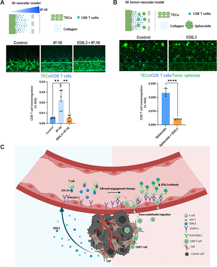
Immune checkpoint blockade (ICB) has become the standard of care for several solid tumors. Multiple combinatorial approaches have been studied to improve therapeutic efficacy. The combination of antiangiogenic agents and ICB has demonstrated efficacy in several cancers. To improve the mechanistic understanding of synergies with these treatment modalities, we performed screens of sera from long-term responding patients treated with ipilimumab and bevacizumab. We discovered a high-titer antibody response against EGF-like repeats and discoidin I-like domains protein 3 (EDIL3) that correlated with favorable clinical outcomes. EDIL3 is an extracellular protein, previously identified as a marker of poor prognosis in various malignancies. Our Tumor Immune Dysfunction and Exclusion analysis predicted that EDIL3 was associated with immune exclusion signatures for cytotoxic immune cell infiltration and nonresponse to ICB. Cancer-associated fibroblasts (CAF) were predicted as the source of EDIL3 in immune exclusion-related cells. Furthermore, The Cancer Genome Atlas Skin Cutaneous Melanoma (TCGA-SKCM) and CheckMate 064 data analyses correlated high levels of EDIL3 with increased pan-fibroblast TGFβ response, enrichment of angiogenic signatures, and induction of epithelial-to-mesenchymal transition. Our in vitro studies validated EDIL3 overexpression and TGFβ regulation in patient-derived CAFs. In pretreatment serum samples from patients, circulating levels of EDIL3 were associated with circulating levels of VEGF, and like VEGF, EDIL3 increased the angiogenic abilities of patient-derived tumor endothelial cells (TEC). Mechanistically, three-dimensional microfluidic cultures and two-dimensional transmigration assays with TEC endorsed EDIL3-mediated disruption of the lymphocyte function-associated antigen-1 (LFA-1)-ICAM-1 interaction as a possible means of T-cell exclusion. We propose EDIL3 as a potential target for improving the transendothelial migration of immune cells and efficacy of ICB therapy.


Figures

Figure 1. Humoral immune responses elicited to EDIL3 were associated with clinical outcomes in patients with metastatic melanoma receiving ipilimumab plus bevacizumab (Ipi-Bev). A, EDIL3-specific antibody FC measured by ELISA in posttreatment versus pretreatment plasma samples of 42 Ipi-Bev patients. CR/PR (green); SD, stable disease (blue); and PD, progressive disease (red). FC ≥1.5 considered clinically significant. B, Frequencies of EDIL3-specific antibody (FC ≥1.5) by clinical responses. C, Immunoblot analysis of EDIL3-specific Ig expression in pretreatment (Pre) and posttreatment (Post) patient's plasma samples of responders. Polyclonal anti-EDIL3 (Abcam) was used in the first lane. Densitometric quantification analysis for relative band intensities shown. D, Kaplan–Meier survival analyses for patients with FC ≥1.5 or <1.5 for EDIL3-specific antibody to Ipi-Bev treatment. E, Percentage of patients with EDIL3-specific antibody FC ≥1.5 in patient cohorts treated with Ipi-Bev (n = 42), ipilimumab (n = 34), PD-1 blockade (n = 21), and nivolumab-ipilimumab (n = 41) therapy.
Figure 1.
Humoral immune responses elicited to EDIL3 were associated with clinical outcomes in patients with metastatic melanoma receiving ipilimumab plus bevacizumab (Ipi-Bev). A, EDIL3-specific antibody FC measured by ELISA in posttreatment versus pretreatment plasma samples of 42 Ipi-Bev patients. CR/PR (green); SD, stable disease (blue); and PD, progressive disease (red). FC ≥1.5 considered clinically significant. B, Frequencies of EDIL3-specific antibody (FC ≥1.5) by clinical responses. C, Immunoblot analysis of EDIL3-specific Ig expression in pretreatment (Pre) and posttreatment (Post) patient's plasma samples of responders. Polyclonal anti-EDIL3 (Abcam) was used in the first lane. Densitometric quantification analysis for relative band intensities shown. D, Kaplan–Meier survival analyses for patients with FC ≥1.5 or <1.5 for EDIL3-specific antibody to Ipi-Bev treatment. E, Percentage of patients with EDIL3-specific antibody FC ≥1.5 in patient cohorts treated with Ipi-Bev (n = 42), ipilimumab (n = 34), PD-1 blockade (n = 21), and nivolumab-ipilimumab (n = 41) therapy.
Figure 2. EDIL3 expression is associated with tumor T-cell immune exclusion gene signatures by TIDE analyses. A, Heat maps showing the correlation (Kendall rank) between EDIL3, MFGE8 and CTNNB1 genes expression and TIDE-predicted signatures for CTL exclusion and dysfunction respectively within TCGA tumor types. B, EDIL3, MFGE8, and CTNNB1 log2(TPM+1) expression and TIDE-predicted value for immunotherapy response in TCGA-SKCM dataset with EDIL3 expression significantly higher among predicted nonresponders (P = 7.4e-11, two-sided Mann–Whitney U test). In the box and whisker plots, the box delineates the first and third quartiles and is bisected by the median; whiskers extend to a maximum of 1.5 times the interquartile range.
Figure 2.
EDIL3 expression is associated with tumor T-cell immune exclusion gene signatures by TIDE analyses. A, Heat maps showing the correlation (Kendall rank) between EDIL3, MFGE8 and CTNNB1 genes expression and TIDE-predicted signatures for CTL exclusion and dysfunction respectively within TCGA tumor types. B,EDIL3, MFGE8, and CTNNB1 log2(TPM+1) expression and TIDE-predicted value for immunotherapy response in TCGA-SKCM dataset with EDIL3 expression significantly higher among predicted nonresponders (P = 7.4e-11, two-sided Mann–Whitney U test). In the box and whisker plots, the box delineates the first and third quartiles and is bisected by the median; whiskers extend to a maximum of 1.5 times the interquartile range.
Figure 3. EDIL3 expression associated with TGFβ signaling, EMT, and angiogenesis signatures. A and B, EDIL3 expression correlated with core biological pathways. EDIL3 expression ordered from low (left) to high (right). Rows display gene expression (z scores normalized) for genes grouped under pathway expression for TGFβ signaling, TGFβ signaling in fibroblast, EMT and angiogenesis signatures in TCGA-SKCM (n = 469; A) and CheckMate 064 (n = 90; B) databases. C, EDIL3 expression correlated with CAF FAP signature score using TIDE.
Figure 3.
EDIL3 expression associated with TGFβ signaling, EMT, and angiogenesis signatures. A and B,EDIL3 expression correlated with core biological pathways. EDIL3 expression ordered from low (left) to high (right). Rows display gene expression (z scores normalized) for genes grouped under pathway expression for TGFβ signaling, TGFβ signaling in fibroblast, EMT and angiogenesis signatures in TCGA-SKCM (n = 469; A) and CheckMate 064 (n = 90; B) databases. C,EDIL3 expression correlated with CAF FAP signature score using TIDE.
Figure 4. EDIL3 is abundantly expressed in CAFs and is upregulated by TGFβ. A, Detection by ELISA of secreted EDIL3 in conditioned medium from NF and patient-derived CAFs (P4-CAF and CAF2). B and C, NF pretreated with or without LY2109761 (LY) followed by TGFβ treatment for 24 hours. EDIL3 expression was examined by B, ELISA of conditioned medium and C, immunoblot analyses of whole-cell lysates. D, qRT-PCR analysis of EDIL3 silencing by siRNA in P4-CAF. EDIL3 mediated regulation of TGFβ target genes Transgelin (TAGLN; E) and α-SMA (ACTA2; F). G, Immunoblot analysis of EDIL3 expression in P4-CAFs treated with VEGF-A (20 ng/mL) or/and bevacizumab (25 μg/mL) for 24 hours. β-actin was used as a loading control. All results are presented as means ± SD and represent three independent experiments. Statistical significance was determined by t test and indicated by P values or as **, P < 0.01; ****, P < 0.0001.
Figure 4.
EDIL3 is abundantly expressed in CAFs and is upregulated by TGFβ. A, Detection by ELISA of secreted EDIL3 in conditioned medium from NF and patient-derived CAFs (P4-CAF and CAF2). B and C, NF pretreated with or without LY2109761 (LY) followed by TGFβ treatment for 24 hours. EDIL3 expression was examined by B, ELISA of conditioned medium and C, immunoblot analyses of whole-cell lysates. D, qRT-PCR analysis of EDIL3 silencing by siRNA in P4-CAF. EDIL3 mediated regulation of TGFβ target genes Transgelin (TAGLN;E) and α-SMA (ACTA2;F). G, Immunoblot analysis of EDIL3 expression in P4-CAFs treated with VEGF-A (20 ng/mL) or/and bevacizumab (25 μg/mL) for 24 hours. β-actin was used as a loading control. All results are presented as means ± SD and represent three independent experiments. Statistical significance was determined by t test and indicated by P values or as **, P < 0.01; ****, P < 0.0001.
Figure 5. Circulating EDIL3 expression correlates with serum VEGF levels and angiogenesis signatures in TCGA-SKCM and CheckMate 064 databases. A, Spearman rank correlation analysis of pretreatment circulating serum levels of EDIL3 versus VEGF-A in Ipi-Bev–treated patients with melanoma (n = 39). B, EDIL3 log2(TPM+1) transformed expression is significantly associated with high angiogenic signatures (Angio) in TCGA-SKCM and CheckMate 064 datasets (two-sided Mann–Whitney U test). C and D, EDIL3 (50 ng/mL) promoted migration of patient-derived endothelial cells similar to VEGF-A (20 ng/mL) assessed by wound healing assay. E–G, EDIL3 promoted tube formation ability of patient-derived endothelial cells similar to VEGF-A. All results are presented as means ± SD and represent three independent experiments. Statistical significance indicated by P values or as *, P < 0.05.
Figure 5.
Circulating EDIL3 expression correlates with serum VEGF levels and angiogenesis signatures in TCGA-SKCM and CheckMate 064 databases. A, Spearman rank correlation analysis of pretreatment circulating serum levels of EDIL3 versus VEGF-A in Ipi-Bev–treated patients with melanoma (n = 39). B,EDIL3 log2(TPM+1) transformed expression is significantly associated with high angiogenic signatures (Angio) in TCGA-SKCM and CheckMate 064 datasets (two-sided Mann–Whitney U test). C and D, EDIL3 (50 ng/mL) promoted migration of patient-derived endothelial cells similar to VEGF-A (20 ng/mL) assessed by wound healing assay. E–G, EDIL3 promoted tube formation ability of patient-derived endothelial cells similar to VEGF-A. All results are presented as means ± SD and represent three independent experiments. Statistical significance indicated by P values or as *, P < 0.05.
Figure 6. EDIL3 blocks immune–endothelial cell adhesion and inhibits T-cell migration. A, Expression of LFA-1(CD11a) on immune cells by flow cytometry. B, Schematic representation of immune–endothelial cell adhesion assay showing EDIL3 antagonizes LFA-1/ICAM-1–dependent adhesion. Graphics created with Biorender. C, rEDIL3 (10, 50, and 200 ng/mL) inhibits adhesion of activated T cells to TNFα-activated tumor endothelial cell's monolayer. D, rEDIL3 (50 ng/mL) blocks the immune–endothelial cell adhesion by interfering with the LFA-1 and ICAM-1 interaction. E, EDIL3 silencing in endothelial cells potentiated T-cell transendothelial migration. F, rEDIL3 partially blocked T-cell transendothelial migration induced by IP-10. All results are presented as means ± SD and represent three independent experiments. Statistical significance was determined by t test and indicated by P values or as *, P < 0.05; **, P < 0.01.
Figure 6.
EDIL3 blocks immune–endothelial cell adhesion and inhibits T-cell migration. A, Expression of LFA-1(CD11a) on immune cells by flow cytometry. B, Schematic representation of immune–endothelial cell adhesion assay showing EDIL3 antagonizes LFA-1/ICAM-1–dependent adhesion. Graphics created with Biorender. C, rEDIL3 (10, 50, and 200 ng/mL) inhibits adhesion of activated T cells to TNFα-activated tumor endothelial cell's monolayer. D, rEDIL3 (50 ng/mL) blocks the immune–endothelial cell adhesion by interfering with the LFA-1 and ICAM-1 interaction. E,EDIL3 silencing in endothelial cells potentiated T-cell transendothelial migration. F, rEDIL3 partially blocked T-cell transendothelial migration induced by IP-10. All results are presented as means ± SD and represent three independent experiments. Statistical significance was determined by t test and indicated by P values or as *, P < 0.05; **, P < 0.01.
Figure 7. EDIL3 blocks transendothelial CD8+ T-cell migration in microfluidic 3D cocultures. Representative images and quantification of migrated CD8+ T-cell numbers in adjacent extracellular matrix (region of interest) after 48 hours of coculture with, vasculature from patient-derived endothelial cells in the presence of the chemoattractant IP-10 (A) or tumor spheroids as the source of IP-10 with vasculature from patient-derived endothelial cells (B). rEDIL3 (50 ng/mL) pretreatment of CD8+ T cells blocked their transendothelial migration. All results are presented as means ± SD and represent three independent experiments. Statistical significance was determined by t test and indicated by P values or as **, P < 0.01; ****, P < 0.0001. C, Model of effects for anti-EDIL3 humoral responses in patients with advanced cancer: Tumor-derived EDIL3 mediates T-cell exclusion in the TME. EDIL3 binds to LFA-1 on T cells and prevents their adherence via ICAM-1 on TECs, thus impeding immune cell extravasation into the TME. ICB may augment a humoral response against EDIL3 accompanied by activated tumor endothelium and increased CD8+ T-cell infiltration. Patients with melanoma with anti-EDIL3 humoral immune responses demonstrated improved therapeutic response to treatment. In addition, immunosuppressive CAFs were identified as an alternate source for EDIL3 in the TME. Graphics created with Biorender.
Figure 7.
EDIL3 blocks transendothelial CD8+ T-cell migration in microfluidic 3D cocultures. Representative images and quantification of migrated CD8+ T-cell numbers in adjacent extracellular matrix (region of interest) after 48 hours of coculture with, vasculature from patient-derived endothelial cells in the presence of the chemoattractant IP-10 (A) or tumor spheroids as the source of IP-10 with vasculature from patient-derived endothelial cells (B). rEDIL3 (50 ng/mL) pretreatment of CD8+ T cells blocked their transendothelial migration. All results are presented as means ± SD and represent three independent experiments. Statistical significance was determined by t test and indicated by P values or as **, P < 0.01; ****, P < 0.0001. C, Model of effects for anti-EDIL3 humoral responses in patients with advanced cancer: Tumor-derived EDIL3 mediates T-cell exclusion in the TME. EDIL3 binds to LFA-1 on T cells and prevents their adherence via ICAM-1 on TECs, thus impeding immune cell extravasation into the TME. ICB may augment a humoral response against EDIL3 accompanied by activated tumor endothelium and increased CD8+ T-cell infiltration. Patients with melanoma with anti-EDIL3 humoral immune responses demonstrated improved therapeutic response to treatment. In addition, immunosuppressive CAFs were identified as an alternate source for EDIL3 in the TME. Graphics created with Biorender.

References

    1. Olbryt M, Rajczykowski M, Widlak W. Biological factors behind melanoma response to immune checkpoint inhibitors. Int J Mol Sci 2020;21:4071. - PMC - PubMed
    1. Zhu L, Yu X, Wang L, Liu J, Qu Z, Zhang H, et al. . Angiogenesis and immune checkpoint dual blockade in combination with radiotherapy for treatment of solid cancers: opportunities and challenges. Oncogenesis 2021;10:47. - PMC - PubMed
    1. Zhang Y, Huang H, Coleman M, Ziemys A, Gopal P, Kazmi SM, et al. . VEGFR2 activity on myeloid cells mediates immune suppression in the tumor microenvironment. JCI Insight 2021;6:e150735. - PMC - PubMed
    1. Hodi FS, Lawrence D, Lezcano C, Wu X, Zhou J, Sasada T, et al. . Bevacizumab plus ipilimumab in patients with metastatic melanoma. Cancer Immunol Res 2014;2:632–42. - PMC - PubMed
    1. Wu X, Giobbie-Hurder A, Connolly EM, Li J, Liao X, Severgnini M, et al. . Anti-CTLA-4 based therapy elicits humoral immunity to galectin-3 in patients with metastatic melanoma. Oncoimmunology 2018;7:e1440930. - PMC - PubMed

Publication types

MeSH terms

Substances