. 2023 Nov 6;155(11):e202313418.
doi: 10.1085/jgp.202313418. Epub 2023 Sep 20.

Mechanisms of dihydropyridine agonists and antagonists in view of cryo-EM structures of calcium and sodium channels

Affiliations

Mechanisms of dihydropyridine agonists and antagonists in view of cryo-EM structures of calcium and sodium channels

Denis B Tikhonov et al. J Gen Physiol. .

Abstract

Opposite effects of 1,4-dihydropyridine (DHP) agonists and antagonists on the L-type calcium channels are a challenging problem. Cryo-EM structures visualized DHPs between the pore-lining helices S6III and S6IV in agreement with published mutational data. However, the channel conformations in the presence of DHP agonists and antagonists are virtually the same, and the mechanisms of the ligands' action remain unclear. We docked the DHP agonist S-Bay k 8644 and antagonist R-Bay k 8644 in Cav1.1 channel models with or without π-bulges in helices S6III and S6IV. Cryo-EM structures of the DHP-bound Cav1.1 channel show a π-bulge in helix S6III but not in S6IV. The antagonist's hydrophobic group fits into the hydrophobic pocket formed by residues in S6IV. The agonists' polar NO2 group is too small to fill up the pocket. A water molecule could sterically fit into the void space, but its contacts with isoleucine in helix S6IV (motif INLF) would be unfavorable. In a model with π-bulged S6IV, this isoleucine turns away from the DHP molecule and its position is occupied by the asparagine from the same motif INLF. The asparagine provides favorable contacts for the water molecule at the agonist's NO2 group but unfavorable contacts for the antagonist's methoxy group. In our models, the DHP antagonist stabilizes entirely α-helical S6IV. In contrast, the DHP agonist stabilizes π-bulged helix S6IV whose C-terminal part turned and rearranged the activation-gate region. This would stabilize the open channel. Thus, agonists, but not antagonists, would promote channel opening by stabilizing π-bulged helix S6IV.


Conflict of interest statement

Disclosures: The authors declare no competing interests exist.

Figures
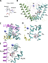
Figure 1.
Figure 1.
DHPs in complexes with calcium channels. (A) Structure of the DHP antagonist R-Bay k 8644. The dihydropyridine ring has a flattened-boat conformation with the polar group NO2 at the starboard, the hydrophobic group COOCH3 at the portside, and phenyl-trifluoromethyl group at the bowsprit. (B) Localization of the DHP binding site in the interface between repeats III and IV. The selectivity filter (SF) and the activation gate regions are marked. (C and D) Intracellular (C) and membrane (D) views of 3-D-aligned structures of DHP-bound calcium channels (PDB IDs: 6jp5, 6jp8, 7jpl, and 7jpw). The backbone helices are shown only for the Cav1.1 channel with R-Bay k 8644 (7jpw). Repeats I, II, III, and IV are colored green, yellow, cyan, and magenta, respectively. A calcium ion in the SF region is shown as a blue sphere. (E) Structure of the Cav1.1 channel with the DHP antagonist nifedipine (6jp5) bound in the fenestration between repeats III and IV. Surrounding residues from helices P1III, S6III, and S6IV are shown as thin sticks.
Figure 2.
Figure 2.
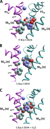
Interactions of DHP ligands with helix S6IV in MC-minimized structures. (A) The portside methoxy group of antagonist R-Bay k 8644 fits in the hydrophobic pocket between Ala4i15 and Ile4i19 (7jpw-based model). (B) A small NO2 group of agonist S-Bay k 8644 does not fill up the pocket, leaving a void space (7jpl-based model). (C) The void space in the 7jpl-based model is filled up by a water molecule that donates two H-bonds to the NO2 group of the agonist.
Figure 3.
Figure 3.
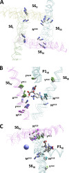
Different orientation of CA–CB bonds in calcium and sodium channels with or without π-bulges in S6 helices. Shown are superimposed structures 7jpw (πaπa), 7xm9 (πaππ), 6jpa (πααα), 6kzp (ππαα), 5x0m (ππππ), 7fbs (παπα), and 7w9p (παππ). (A) Cytoplasmic view of CA–CB bonds in positions i20. In the π-bulged helices S6, the CA–CB bonds are directed toward the neighboring repeat. In the entirely α-helical S6s, the CA–CB bonds are directed away from the pore lumen. (B and C) Antagonist R-Bay k 8644 in the S6III/S6IV interface of the superimposed structures. Sticks indicate CA–CB bonds in positions of DHP-sensing residues or their homologs in sodium channels. Orientations of the CA–CB bonds in positions 3p44, 3i10, 4i11, and 4i15 are conserved. Orientations of CA–CB bonds in positions 3i18, 4i19, and 4i20 strongly depend on the presence or absence of a π-bulge.
Figure 4.
Figure 4.
Structural diversity of DHP–channel models based on different cryo-EM templates (PDB IDs: 7jpl, 7jpw, 7xm9, 6jpa, 6kzp, 5x0m, 7fbs, and 7w9p). (A) Superimposed MC-minimized models of the Cav1.1 channel with R-Bay k 8644. Backbones and side chains are shown for the Cav1.1 channel with antagonist R-Bay k 8644 in nanodiscs (7jpw). (B) Superimposed MC-minimized models of the Cav1.1 channel with agonist S-Bay k 8644. Backbones and side chains are shown for the model based on sodium channel template 7w9p. Small red spheres indicate the positions of a water molecule. (C) Various orientations of Met3i19 in the Cav1.1 channel models with antagonist R-Bay k 8644. The methionine side chain interacts with the agonist in models with π-bulged, but not entirely α-helical S6III. (D) Various orientations of asparagine N4i20 in the channel models with agonist S-Bay k 8644. The asparagine side chain interacts with the ligand in models with π-bulged, but not entirely α-helical S6IV. In C and D, examples of both α-helical and π-bulged S6III and S6IV are shown as α-tracing with different colors.
Figure 5.
Figure 5.
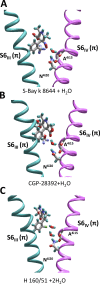
Models of the Cav1.1 channel with agonists bound to π-bulged S6. (A) Model with agonist S-Bay k 8644 obtained with constraint-driven approach. A water molecule donates an H-bond to the agonist polar portside group and another H-bond to the backbone CO group of Ala4i15. The side chain of Asn4i20 donates an H-bond to the CO group of Phe4i16 and another H-bond to the water molecule. The backbone CO groups in positions 4i15 and 4i16 are bachelors in the π-bulged helix S6IV. Thus, ligand–channel interactions stabilize the π-bulged helix S6IV. (B) Model with agonist S-202-791. Ligand–water–channel interactions are very similar to those shown in A. (C) Model with agonist H 160/51, which lacks a portside group. Two water molecules were imposed to fill the gap between the agonist and helix S6IV.
Figure 6.
Figure 6.
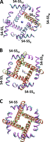
α–π Transition in S6IV and the pore dimensions. (A) Cytoplasmic view at the superimposition of the 7fbs-based model (magenta, red S6IV) and the resulting model obtained by constraint-driven α to π transition (cyan, green S6IV). S-Bay k 8644 is shown as sticks and a calcium ion as a blue sphere. The C-end of S6IV is slightly shifted toward the pore axis. (B) Superimposition of the Nav1.7 apo state 7w9k (magenta, red S6) and the channel complex with bupivacaine 8i5b (cyan, green S6s). Bupivacaine is shown as sticks. In the apo state, S6IV is in the α-form; binding of the drug causes S6IV transition to the π-form. The cuff formed by helices S4–S5 is the same in both structures and it does not allow the S6 helices to diverge. As a result, disruption of intersegment contacts by α to π transition can cause only inward movement of S6s. (C) Open (6dvz, cyan S4–S5, green S6) and closed (6mho, magenta S4–S5, red S6) structures of TRPV3. In the closed-pore structure, segments S6 are entirely α-helical and in the open-pore structure they have a π-helical turn. Note that in the open structure, both S4–S5 and S6 cooperatively moved outward the pore axis as compared with the closed structure.
Figure 7.
Figure 7.
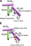
The proposed scheme of action of DHP antagonists and agonists. (A) The antagonist binding stabilizes entirely the α-helical conformation of S6IV and favorable interaction of the hydrophobic portside group with Ile4i19. (B) The agonist stabilizes a π-bulged S6IV by polar contacts with N4i20 via a water molecule (red sphere). The α and π forms of S6IV drastically differ by the orientation of residues at the C-end of the helix (Ile4i26 is shown as an example). The altered orientation switches contacts with neighboring segments (linker-helix S4–S5IV and helix S6I) at the gate region. Different patterns of these contacts can specifically stabilize the open (agonist) or closed (antagonist) states of the pore domain.

Comment in

Similar articles

Cited by

References

    1. Brooks, C.L., III, Pettitt M., and Karplus M.. 1985. Structural and energetic effects of truncating long ranged interactions in ionic and polar fluids. J. Chem. Phys. 83:5897–5908. 10.1063/1.449621 - DOI
    1. Brown, A.M., Kunze D.L., and Yatani A.. 1984. The agonist effect of dihydropyridines on Ca channels. Nature. 311:570–572. 10.1038/311570a0 - DOI - PubMed
    1. Catterall, W.A. 2011. Voltage-gated calcium channels. Cold Spring Harb. Perspect. Biol. 3:a003947. 10.1101/cshperspect.a003947 - DOI - PMC - PubMed
    1. Catterall, W.A., Lenaeus M.J., and Gamal El-Din T.M.. 2020. Structure and pharmacology of voltage-gated sodium and calcium channels. Annu. Rev. Pharmacol. Toxicol. 60:133–154. 10.1146/annurev-pharmtox-010818-021757 - DOI - PubMed
    1. Choudhury, K., and Delemotte L.. 2023. Modulation of pore opening of eukaryotic sodium channels by π-helices in S6. J. Phys. Chem. Lett. 14:5876–5881. 10.1021/acs.jpclett.3c00803 - DOI - PMC - PubMed

MeSH terms