

Semaphorins: Missing Signals in Age-dependent Alteration of Neuromuscular Junctions and Skeletal Muscle Regeneration
- PMID: 37728580
- PMCID: PMC10917540
- DOI: 10.14336/AD.2023.0801
Semaphorins: Missing Signals in Age-dependent Alteration of Neuromuscular Junctions and Skeletal Muscle Regeneration
Abstract
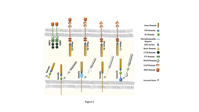
Skeletal muscle is characterized by a remarkable capacity to rearrange after physiological changes and efficiently regenerate. However, during aging, extensive injury, or pathological conditions, the complete regenerative program is severely affected, with a progressive loss of muscle mass and function, a condition known as sarcopenia. The compromised tissue repair program is attributable to the gradual depletion of stem cells and to altered regulatory signals. Defective muscle regeneration can severely affect re-innervation by motor axons, and neuromuscular junctions (NMJs) development, ultimately leading to skeletal muscle atrophy. Defects in NMJ formation and maintenance occur physiologically during aging and are responsible for the pathogenesis of several neuromuscular disorders. However, it is still largely unknown how neuromuscular connections are restored on regenerating fibers. It has been suggested that attractive and repelling signals used for axon guidance could be implicated in this process; in particular, guidance molecules called semaphorins play a key role. Semaphorins are a wide family of extracellular regulatory signals with a multifaceted role in cell-cell communication. Originally discovered as axon guidance factors, they have been implicated in cancer progression, embryonal organogenesis, skeletal muscle innervation, and other physiological and developmental functions in different tissues. In particular, in skeletal muscle, specific semaphorin molecules are involved in the restoration and remodeling of the nerve-muscle connections, thus emphasizing their plausible role to ensure the success of muscle regeneration. This review article aims to discuss the impact of aging on skeletal muscle regeneration and NMJs remodeling and will highlight the most recent insights about the role of semaphorins in this context.
Conflict of interest statement
We declare no conflicts of interest.
Figures


Similar articles
-
Inducible depletion of adult skeletal muscle stem cells impairs the regeneration of neuromuscular junctions.Elife. 2015 Aug 27;4:e09221. doi: 10.7554/eLife.09221. Elife. 2015. PMID: 26312504 Free PMC article.
-
Muscle Fibers Secrete FGFBP1 to Slow Degeneration of Neuromuscular Synapses during Aging and Progression of ALS.J Neurosci. 2017 Jan 4;37(1):70-82. doi: 10.1523/JNEUROSCI.2992-16.2016. J Neurosci. 2017. PMID: 28053031 Free PMC article.
-
Older mice show decreased regeneration of neuromuscular junctions following lengthening contraction-induced injury.Geroscience. 2023 Jun;45(3):1899-1912. doi: 10.1007/s11357-023-00774-w. Epub 2023 Mar 23. Geroscience. 2023. PMID: 36952126 Free PMC article.
-
The emerging role of the sympathetic nervous system in skeletal muscle motor innervation and sarcopenia.Ageing Res Rev. 2021 May;67:101305. doi: 10.1016/j.arr.2021.101305. Epub 2021 Feb 18. Ageing Res Rev. 2021. PMID: 33610815 Free PMC article. Review.
-
The Composition, Development, and Regeneration of Neuromuscular Junctions.Curr Top Dev Biol. 2018;126:99-124. doi: 10.1016/bs.ctdb.2017.08.005. Epub 2017 Nov 10. Curr Top Dev Biol. 2018. PMID: 29305005 Review.
Cited by
-
Matrisome Transcriptome Dynamics during Tissue Aging.Life (Basel). 2024 May 7;14(5):593. doi: 10.3390/life14050593. Life (Basel). 2024. PMID: 38792614 Free PMC article.
References
-
- Frontera WR, Ochala J (2015). Skeletal muscle: a brief review of structure and function. Calcif Tissue Int, 96:183-195. - PubMed
Publication types
MeSH terms
Substances
LinkOut - more resources
Full Text Sources