

Retinal artery occlusion after facial filler injection in a patient with patent foramen ovale: a case report and literature review
- PMID: 37728598
- PMCID: PMC10515541
- DOI: 10.1177/03000605231194514
Retinal artery occlusion after facial filler injection in a patient with patent foramen ovale: a case report and literature review
Abstract
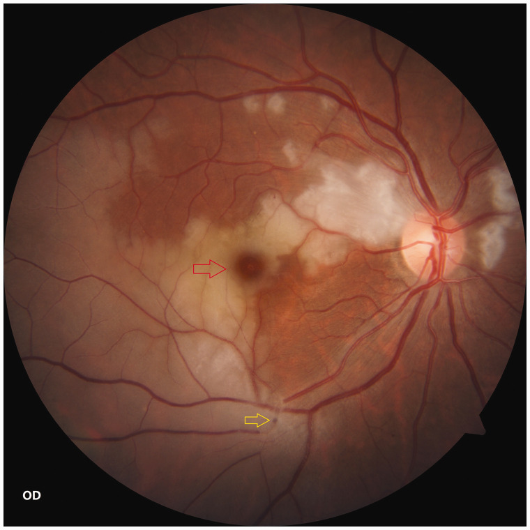
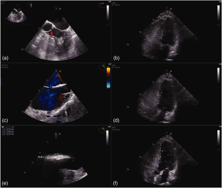
We describe a young woman with patent foramen ovale who developed multiple retinal artery occlusion in the right eye after injection of hyaluronic acid into the nasal root. She reported a gradual decline in visual acuity, with visual field defects that had developed in two stages. Multiple retinal artery occlusion was confirmed by slit-lamp examination, dilated fundus examination, optical coherence tomography, visual field examination, and fundus fluorescein angiography. A patent foramen ovale was detected by electrocardiography, transesophageal echocardiography, and transthoracic sonography. The patient was treated with intravenous dexamethasone and cobamamide, as well as extracorporeal counterpulsation therapy; this approach has not been described in previous literature regarding retinal artery occlusion. The patient's visual acuity improved from counting fingers at 30 cm to 20/133 within 3 days. Our report emphasizes the need for better understanding of vascular anatomy to minimize the risk of complications. Moreover, patients undergoing hyaluronic acid injection should receive information regarding the potential for mild and severe complications; relevant tests should be performed before surgery to exclude vulnerable patients. Finally, a nursing system is needed to facilitate the emergency recognition, triage, and management of retinal artery occlusion.
Keywords: Hyaluronic acid; case report.; extracorporeal counterpulsation therapy; facial fillers; patent foramen ovale; retinal artery occlusion.
Conflict of interest statement
The authors declare that there is no conflict of interest.
Figures


Similar articles
-
Central retinal artery occlusion associated with patent foramen ovale: a case report and literature review.Arq Bras Oftalmol. 2021 Jul 14;84(5):494-498. doi: 10.5935/0004-2749.20210073. eCollection 2021. Arq Bras Oftalmol. 2021. PMID: 34320104 Free PMC article. Review.
-
Central retinal artery occlusion as a result of symptomatic patent foramen ovale.Rom J Ophthalmol. 2023 Jan-Mar;67(1):69-72. doi: 10.22336/rjo.2023.12. Rom J Ophthalmol. 2023. PMID: 37089812 Free PMC article.
-
Monocular central retinal artery occlusion caused by bilateral internal carotid artery hypoplasia complicated with patent foramen ovale: a case report and review of literature.Eur J Med Res. 2021 Jun 13;26(1):55. doi: 10.1186/s40001-021-00530-w. Eur J Med Res. 2021. PMID: 34120645 Free PMC article. Review.
-
Undiagnosed patent foramen ovale as a rare cause for branch retinal artery occlusion.Eur J Ophthalmol. 2015 Jul 30;25(5):e88-90. doi: 10.5301/ejo.5000608. Eur J Ophthalmol. 2015. PMID: 25837644
-
Retinal vascular occlusion in pregnancy: three case reports and a review of the literature.J Med Case Rep. 2022 Apr 21;16(1):167. doi: 10.1186/s13256-022-03369-9. J Med Case Rep. 2022. PMID: 35449024 Free PMC article. Review.
Cited by
-
EYE-CODE Protocol for the Nonophthalmologist for Treatment of Retinal Artery Occlusion After Intra-Arterial Injection of Soft-Tissue Fillers: 2025 Update.J Cosmet Dermatol. 2025 Jul;24(7):e70336. doi: 10.1111/jocd.70336. J Cosmet Dermatol. 2025. PMID: 40631662 Free PMC article.
-
Ocular Manifestations and Complications of Patent Foramen Ovale: A Narrative Review.J Pers Med. 2024 Jun 27;14(7):695. doi: 10.3390/jpm14070695. J Pers Med. 2024. PMID: 39063949 Free PMC article. Review.
-
Congenital heart disease: types, pathophysiology, diagnosis, and treatment options.MedComm (2020). 2024 Jul 5;5(7):e631. doi: 10.1002/mco2.631. eCollection 2024 Jul. MedComm (2020). 2024. PMID: 38974713 Free PMC article. Review.
References
-
- Beleznay K, Carruthers JDA, Humphrey S, et al.. Update on avoiding and treating blindness from fillers: a recent review of the world literature. Aesthet Surg J 2019; 39: 662–674. - PubMed
-
- Crowley JS, Kream E, Fabi S, et al.. Facial rejuvenation with fat grafting and fillers. Aesthet Surg J 2021; 41: S31–S38. - PubMed
-
- Thulesen J. Iatrogenic vision loss following aesthetic treatment with hyaluronic acid-containing filler: every injector should be prepared. Dermatol Ther 2020; 33: e13913. - PubMed
-
- Kapoor KM, Kapoor P, Heydenrych I, et al.. Vision loss associated with hyaluronic acid fillers: a systematic review of literature. Aesthetic Plast Surg 2020; 44: 929–944. - PubMed
Publication types
MeSH terms
Substances
LinkOut - more resources
Full Text Sources
