. 2023 Nov 6;222(11):e202210093.
doi: 10.1083/jcb.202210093. Epub 2023 Sep 20.

UHRF1 promotes spindle assembly and chromosome congression by catalyzing EG5 polyubiquitination

Affiliations

UHRF1 promotes spindle assembly and chromosome congression by catalyzing EG5 polyubiquitination

Xuli Qi et al. J Cell Biol. .

Abstract

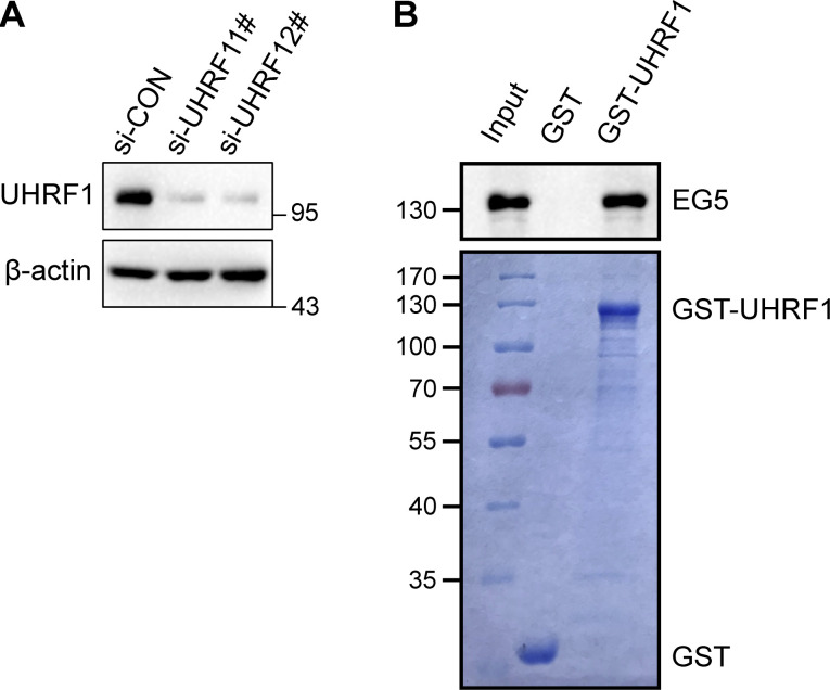
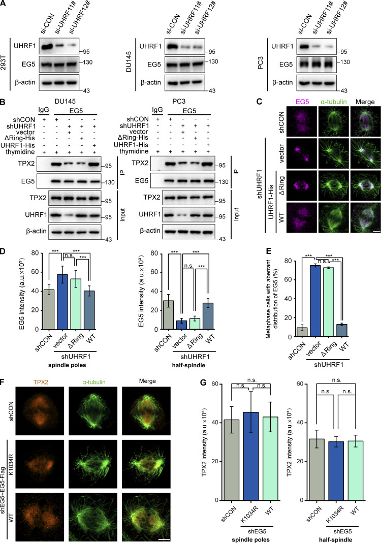
UHRF1 is an epigenetic coordinator bridging DNA methylation and histone modifications. Additionally, UHRF1 regulates DNA replication and cell cycle, and its deletion induces G1/S or G2/M cell cycle arrest. The roles of UHRF1 in the regulation of G2/M transition remain poorly understood. UHRF1 depletion caused chromosome misalignment, thereby inducing cell cycle arrest at mitotic metaphase, and these cells exhibited the defects of spindle geometry, prominently manifested as shorter spindles. Mechanistically, UHRF1 protein directly interacts with EG5, a kinesin motor protein, during mitosis. Furthermore, UHRF1 induced EG5 polyubiquitination at the site of K1034 and further promoted the interaction of EG5 with spindle assembly factor TPX2, thereby ensuring accurate EG5 distribution to the spindles during metaphase. Our study clarifies a novel UHRF1 function as a nuclear protein catalyzing EG5 polyubiquitination for proper spindle architecture and faithful genomic transmission, which is independent of its roles in epigenetic regulation and DNA damage repair inside the nucleus. These findings revealed a previously unknown mechanism of UHRF1 in controlling mitotic spindle architecture and chromosome behavior and provided mechanistic evidence for UHRF1 deletion-mediated G2/M arrest.


Conflict of interest statement

Disclosures: The authors declare no competing interests exist.

Figures

Figure 1.
Figure 1.
Depletion of UHRF1 causes chromosome misalignment. (A and B) DU145 and PC3 cells with or without UHRF1 depletion were established by stable transfection with shRNA vectors for UHRF1 or control, and UHRF1 expression was assessed by Western blotting (A). The cell cycle distribution was analyzed by flow cytometry (B). (C–F) The nuclear DNA in DU145 and PC3 cells with or without UHRF1 depletion was stained with DAPI (scale bar, 20 μm; inset scale bar, 10 μm, C). The percent of mitotic cells was assessed according to nuclei morphology. n > 100 cells (D). The chromosomes that failed to congress at the metaphase plate are highlighted by red arrows (scale bar, 5 μm, E). The percentage of cells with unaligned chromosomes was assessed. n > 80 cells (F). (G and H) DU145 cells with UHRF1 depletion were transiently transfected with plasmids expressing UHRF1ΔRING or UHRF1WT, and the nuclear DNA was stained with DAPI (scale bar, 5 μm, G). The percentage of cells with unaligned chromosomes was assessed. n > 80 cells (H). The data for quantification in D, F, and H are from n = 3 independent experiments. Results are represented as mean ± SD (one-way ANOVA test); error bars represent SD. n.s., not significant; **, P < 0.01; ***, P < 0.001. Source data are available for this figure: SourceData F1.
Figure S1.
Figure S1.
In vitro recombinant proteins GST-pull down assay. (A) Knockdown of UHRF1. DU145 cells were transiently transfected with UHRF1 siRNAs and UHRF1 expression was evaluated by Western blotting. (B) GST-pulldown assay of UHRF1 using the indicated GST fusion proteins. EG5 protein was detected. Source data are available for this figure: SourceData FS1.
Figure 2.
Figure 2.
Depletion of UHRF1 damages spindle architecture. (A) DU145 cells were transiently transfected with or without siUHRF1. The mitotic spindles were stained with immunofluorescent anti-α-tubulin antibody (green) and chromosomes were stained with DAPI. Scale bar, 5 μm. (B–D) Spindle pole distance was measured. n = 40 cells (B). The percentage of cells at the metaphases with abnormal spindle geometry was assessed (C) in DU145 cells when transfected with or without siUHRF1. n > 50 cells for each condition. The asters fluorescence intensity of the metaphase spindle was measured. n = 40 cells for each condition. (E) DU145 cells with UHRF1 depletion were transiently transfected with plasmids expressing UHRF1ΔRING or UHRF1WT. The mitotic spindles were stained with immunofluorescent α-tubulin antibody and chromosomes were stained with DAPI. Scale bar, 5 μm. (F–H) Spindle pole distance was measured (n = 40 cells; F) and the percentage of cells at the metaphases with abnormal spindle geometry was assessed (n > 50 cells; G). The mean fluorescence intensity of asters was analyzed by ImageJ. n = 40 cells (H). The data for quantification in C, D, G, and H are from n = 3 independent experiments. Results are represented as mean ± SD; error bars represent SD. Dots represent individual cell samples in B and F; bars are median ± quartile. n.s., not significant; ***, P < 0.001. One-way ANOVA test.
Figure 3.
Figure 3.
EG5 is a UHRF1-interactive mitotic protein. (A) EG5 was identified as a component of UHRF1-interactive protein complexes by immunoprecipitation coupled with mass spectrometry. UHRF1-interacting protein complexes were immunoprecipitated with anti-Flag antibody in HEK293T cells expressing Flag-tagged UHRF1, and then were eluted with Flag peptide. The UHRF1-interacted protein complexes were separated by SDS-PAGE and a 125-kD electrophoretic band was subjected to mass spectrometry analysis. (B) HEK293T, DU145, and PC3 cells were transfected with plasmids expressing UHRF1-His or EG5-Flag, and the interaction between exogenous UHRF1 and EG5 proteins was validated by immunoprecipitation with antibodies against His or Flag, followed by immunoblotting. (C) The interaction between endogenous UHRF1 and EG5 proteins was validated by immunoprecipitation with antibodies against UHRF1 or EG5, followed by immunoblotting in HEK293T, DU145, and PC3 cells. (D) DU145 cells were synchronized in G1/S phase using double thymidine blocking and then released by culture media. The cells were synchronized in G2/M phases at 9 h after release. The interaction between endogenous UHRF1 and EG5 proteins was validated by immunoprecipitation with antibodies against UHRF1, followed by immunoblotting. (E and F) GST-tagged full-length UHRF1 and individual subdomains were constructed for mapping the EG5-binding region. Purified recombinant proteins of GST-tagged individual domains of UHRF1 were incubated with HEK293T cell lysates in vitro as indicated, followed by immunoblotting with anti-EG5 antibody (F). The lysate of HEK293T cells was used for a positive control. (G and H) GST-tagged full-length EG5 and individual subdomains were constructed for mapping the UHRF1-binding region. Purified recombinant proteins of GST-tagged individual domains of EG5 were incubated with HEK293T cell lysates in vitro as indicated, followed by immunoblotting with anti-UHRF1 antibody (H). The lysate of HEK293T cells was used for a positive control. Source data are available for this figure: SourceData F3.
Figure 4.
Figure 4.
UHRF1 catalyzes the polyubiquitination of EG5 at the site of K1034. (A) HEK293T cells were cotransfected with control or UHRF1 siRNAs, together with HA-ubiquitin and EG5-Flag. The cells then were synchronized at the G2/M phase with a double thymidine block. EG5 protein was immunoprecipitated with anti-Flag antibody and the ubiquitination level of EG5 was assessed with HA antibody. (B) HEK293T cells were cotransfected with plasmids expressing UHRF1-His, HA-ubiquitin, and EG5-Flag, and then the cells were synchronized at the G2/M phase. EG5 protein was immunoprecipitated with anti-Flag antibody and the ubiquitination level of EG5 was assessed with an HA antibody. (C) HEK293T cells were cotransfected with plasmids expressing UHRF1-His or UHRF1ΔRING-His, HA-ubiquitin, and EG5-Flag, and then the cells were synchronized at the G2/M phase. EG5 protein was immunoprecipitated with anti-Flag antibody and the ubiquitination level of EG5 was assessed with an HA antibody. (D) HEK293T cells were cotransfected with control or UHRF1 siRNAs together with HA-ubiquitin and EG5-Flag, and then the cells were synchronized in the G1/S phase using double thymidine blocking and released by culture media. The cells were synchronized in G2/M phases at 9 h after release. EG5 protein was immunoprecipitated with an anti-Flag antibody and the ubiquitination level of EG5 was assessed with HA antibody. (E and G) HEK293T cells were cotransfected with control or UHRF1 siRNAs, together with HA-K48 or HA-K63 and EG5-Flag. The cells then were synchronized at the G2/M phase with a double thymidine block. EG5 protein was immunoprecipitated with an anti-Flag antibody, and the ubiquitination level of EG5 was assessed with an HA antibody. (F and H) HEK293T cells were cotransfected with plasmids expressing UHRF1-His, HA-K48/HA-K63, and EG5-Flag, and then the cells were synchronized at the G2/M phase. EG5 protein was immunoprecipitated with an anti-Flag antibody, and the ubiquitination level of EG5 was assessed with an HA antibody. (I) In vitro ubiquitination assay was performed in the presence of Ub (WT, K48, or K63), E1, E2, EG5, and UHRF1 (WT or ΔRING mutant). The ubiquitination of EG5 was examined with HA antibody. (J) HEK293T cells were cotransfected with plasmids expressing HA-ubiquitin and EG5-Flag with four lysine mutations as shown, and then the cells were synchronized at the G2/M phase. EG5 protein was immunoprecipitated with anti-Flag antibody, and the ubiquitination level of EG5 was assessed with an HA antibody. (K) HEK293T cells were cotransfected with plasmids expressing UHRF1ΔRING-His, HA-ubiquitin, and EG5-Flag with wild type or lysine mutation as shown, and then the cells were synchronized at the G2/M phase. EG5 protein was immunoprecipitated with anti-Flag antibody and the ubiquitination level of EG5 was assessed with an HA antibody. Source data are available for this figure: SourceData F4.
Figure S2.
Figure S2.
UHRF1 catalyzes the K63-linked ubiquitination of EG5 at K1034. (A and B) HEK293T cells were cotransfected with plasmids expressing UHRF1-His or UHRF1ΔRING-His, HA-K48/HA-K63, and EG5-Flag, and then the cells were synchronized at the G2/M phase. EG5 protein was immunoprecipitated with an anti-Flag antibody, and the ubiquitination level of EG5 was assessed with an HA antibody. (C) HEK293T cells were cotransfected with plasmids expressing HA-K63 and EG5-Flag with four lysine mutations as shown, and then the cells were synchronized at the G2/M phase. EG5 protein was immunoprecipitated with an anti-Flag antibody, and the ubiquitination level of EG5 was assessed with an HA antibody. (D) HEK293T cells were cotransfected with plasmids expressing UHRF1ΔRING-His, HA-K63, and EG5-Flag with wild-type or lysine mutation as shown, and then the cells were synchronized at the G2/M phase. EG5 protein was immunoprecipitated with anti-Flag antibody and the ubiquitination level of EG5 was assessed with an HA antibody. Source data are available for this figure: SourceData FS2.
Figure 5.
Figure 5.
UHRF1 regulates EG5 interactions with TPX2 to determine EG5 localization on the spindle. (A) UHRF1 was depleted with siRNAs in DU145 cells. EG5 were stained with immunofluorescent antibodies (red) and the spindle was stained with anti-α-tubulin antibody (green). Scale bar, 5 μm. (B–D) B and C: Corresponding EG5 fluorescence intensity profiles of cells. n = 40 cells for each condition. EG5 fluorescence intensity of the spindle poles (B) or half-spindle (C) was measured, and the percentage of cells at the metaphases with abnormal distribution of EG5 was assessed. n > 50 cells (D). (E) DU145 or PC3 cells were transiently transfected with UHRF1 siRNAs, EG5 protein was immunoprecipitated, and the interacting TPX2 was assessed by immunoblotting. (F) DU145 or PC3 cells were transiently cotransfected with plasmids expressing EG5WT-Flag or EG5K1034R-Flag, EG5 protein was immunoprecipitated with anti-Flag antibody, and the interacting TPX2 was assessed by immunoblotting. (G) DU145 cells with EG5 depletion were transiently transfected with plasmids expressing EG5WT or EG5K1034R. EG5 were stained with immunofluorescent antibodies (red) and the spindle was stained with anti-α-tubulin antibody (green). Scale bar, 5 μm. (H–J) H and I: Corresponding EG5 fluorescence intensity profiles of cells. n = 40 cells for each condition. EG5 fluorescence intensity of the spindle poles (H) or half-spindle (I) was measured, and the percentage of cells at the metaphases with abnormal distribution of EG5 was assessed. n > 50 cells (J). (K and L) DU145 cells with UHRF1 depletion were transiently transfected with plasmids expressing EG5WT-Flag (K), or DU145 cells were transiently transfected with plasmids expressing Flag-tagged wild-type EG5 (WT)/K1034R mutant (L), and immunoprecipitated with a Flag antibody. Half of the immunoprecipitate was used for ATPase assay (bar graph) and the other half was separated by SDS-PAGE and immunoblotted with Flag antibody (gel image). NT, non-transfected. The data for quantification in B–D and H–L are from n = 3 independent experiments. Results are represented as mean ± SD (one-way ANOVA test); error bars represent SD. *, P < 0.05; ***, P < 0.001. Source data are available for this figure: SourceData F5.
Figure S3.
Figure S3.
EG5 localization and its interaction with TPX2 are affected by UHRF1 mutation. (A) HEK293T, DU145, or PC3 cells were transiently transfected with UHRF1 siRNAs, and UHRF1 or EG5 expression was evaluated by Western blotting. (B) DU145 or PC3 cells with UHRF1 depletion were transiently transfected with plasmids expressing UHRF1ΔRING or UHRF1WT, EG5 protein was immunoprecipitated, and the interacting TPX2 was assessed by immunoblotting. (C) DU145 cells with UHRF1 depletion were transiently transfected with plasmids expressing UHRF1ΔRING or UHRF1WT. EG5 was stained with immunofluorescent antibodies (red), and the spindle was stained with anti-α-tubulin antibody (green). Scale bar, 5 μm. (D and E) Corresponding EG5 fluorescence intensity profiles of cells. n = 40 cells for each condition. EG5 fluorescence intensity of the spindle poles or half-spindle was measured (D), and the percent of cells at the metaphases with abnormal distribution of EG5 was assessed. n > 50 cells (E). (F) DU145 cells with EG5 depletion were transiently transfected with plasmids expressing EG5WT or EG5K1034R. TPX2 were stained with immunofluorescent antibodies (yellow), and the spindle was stained with anti-α-tubulin antibody (green). Scale bar, 5 μm. (G) Corresponding TPX2 fluorescence intensity profiles of cells. n = 40 cells for each condition. TPX2 fluorescence intensity of the spindle poles or half-spindle was measured. The data for quantification in D, E, and G are from n = 3 independent experiments. Results are represented as mean ± SD (one-way ANOVA test); error bars represent SD. n.s., not significant; ***, P < 0.001. Source data are available for this figure: SourceData FS3.
Figure 6.
Figure 6.
UHRF1 ubiquitination is critical for spindle architecture. (A) DU145 cells with EG5 depletion were transiently transfected with plasmids expressing EG5WT or EG5K1034R, and the nuclear DNA was stained with DAPI. Scale bar, 20 μm; inset scale bar, 10 μm. (B) The mitotic cells were identified according to nuclei morphology and the percentage of mitotic cells was assessed. n > 100 cells. (C) Chromosomes that failed to congress at the metaphase plate are highlighted by red arrows. Scale bar, 5 μm. (D) The percentage of DU145 cells with unaligned chromosomes was assessed. n > 50 cells. (E) Spindles were stained with immunofluorescent α-tubulin antibody and chromosomes were stained with DAPI. Scale bar, 5 μm. (F and G) Spindle pole distance was measured. n = 40 cells (F). The percent of cells at the metaphases with abnormal spindle geometry was assessed. n > 50 cells (G). The data for quantification in B, D, and G are from n = 3 independent experiments. Results are represented as mean ± SD; error bars represent SD. Dots represent individual cell samples in F; bars are median ± quartile. **, P < 0.01; ***, P < 0.001. One-way ANOVA test.
Figure 7.
Figure 7.
A schematic model showing the molecular mechanism by which UHRF1 promotes spindle assembly and chromosome congression in mitosis by catalyzing mitotic kinesin motor EG5 polyubiquitination for the interaction with TPX2.

Similar articles

Cited by

References

    1. Acar, S., Carlson D.B., Budamagunta M.S., Yarov-Yarovoy V., Correia J.J., Niñonuevo M.R., Jia W., Tao L., Leary J.A., Voss J.C., et al. . 2013. The bipolar assembly domain of the mitotic motor kinesin-5. Nat. Commun. 4:1343. 10.1038/ncomms2348 - DOI - PMC - PubMed
    1. Alfonso-Pérez, T., Hayward D., Holder J., Gruneberg U., and Barr F.A.. 2019. MAD1-dependent recruitment of CDK1-CCNB1 to kinetochores promotes spindle checkpoint signaling. J. Cell Biol. 218:1108–1117. 10.1083/jcb.201808015 - DOI - PMC - PubMed
    1. Arima, Y., Hirota T., Bronner C., Mousli M., Fujiwara T., Niwa S., Ishikawa H., and Saya H.. 2004. Down-regulation of nuclear protein ICBP90 by p53/p21Cip1/WAF1-dependent DNA-damage checkpoint signals contributes to cell cycle arrest at G1/S transition. Genes Cells. 9:131–142. 10.1111/j.1356-9597.2004.00710.x - DOI - PubMed
    1. Ashraf, W., Ibrahim A., Alhosin M., Zaayter L., Ouararhni K., Papin C., Ahmad T., Hamiche A., Mély Y., Bronner C., and Mousli M.. 2017. The epigenetic integrator UHRF1: On the road to become a universal biomarker for cancer. Oncotarget. 8:51946–51962. 10.18632/oncotarget.17393 - DOI - PMC - PubMed
    1. Ben-David, U., and Amon A.. 2020. Context is everything: Aneuploidy in cancer. Nat. Rev. Genet.. 21:44–62. 10.1038/s41576-019-0171-x - DOI - PubMed

Publication types