. 2023 Dec 1;34(13):ar125.
doi: 10.1091/mbc.E23-06-0248. Epub 2023 Sep 20.

Exploring the "misfolding problem" by systematic discovery and analysis of functional-but-degraded proteins

Affiliations

Exploring the "misfolding problem" by systematic discovery and analysis of functional-but-degraded proteins

Matthew P Flagg et al. Mol Biol Cell. .

Abstract

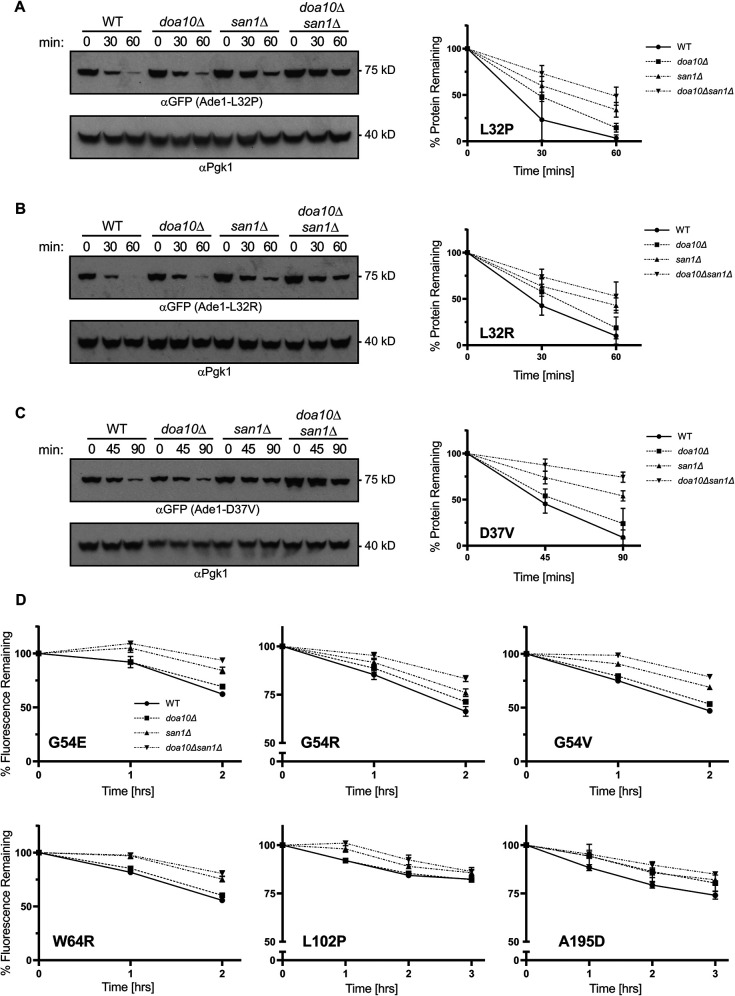
In both health and disease, the ubiquitin-proteasome system (UPS) degrades point mutants that retain partial function but have decreased stability compared with their wild-type counterparts. This class of UPS substrate includes routine translational errors and numerous human disease alleles, such as the most common cause of cystic fibrosis, ΔF508-CFTR. Yet, there is no systematic way to discover novel examples of these "minimally misfolded" substrates. To address that shortcoming, we designed a genetic screen to isolate functional-but-degraded point mutants, and we used the screen to study soluble, monomeric proteins with known structures. These simple parent proteins yielded diverse substrates, allowing us to investigate the structural features, cytotoxicity, and small-molecule regulation of minimal misfolding. Our screen can support numerous lines of inquiry, and it provides broad access to a class of poorly understood but biomedically critical quality-control substrates.


Figures

FIGURE 1:
FIGURE 1:
The position of minimally misfolded mutants in the Ade1 structure. (A) Two perspectives of the Ade1 crystal structure (PDB 1A48). Arrows and color-coded sidechains indicate positions at which destabilizing mutations were isolated. (B–G) Closeups of positions at which destabilizing substitutions were isolated. In each inset, the position’s wild-type amino acid is shown, color coded, and indicated by an arrow. To demonstrate proximity and spatial relationships, additional screen-isolated residues that fall within an inset’s field of view are shown and color coded as in A. In the case of buried positions (A and D–G), additional amino acids that fall within a 5 Å zone are shown in khaki. In the case of intramolecular hydrogen bonding (B), black lines show hydrogen bonds predicted by UCSF Chimera under relaxed constraints.
FIGURE 2:
FIGURE 2:
Ade1 mutants are degraded by San1 and Doa10 in parallel. (A–C) WT, san1Δ, doa10Δ, and san1Δdoa10Δ null strains expressing Ade1-L32P (A), L32R (B), or D37V (C) were grown into log phase and treated with CHX. At the indicated timepoints, cells were harvested and lysed. Lysates were then Western blotted using α-GFP and α-Pgk1. Densitometry was performed with FIJI. Each α-GFP band was normalized to its corresponding α-Pgk1 band, and Pgk1-normalized readings were normalized to t = 0. Graphs show the mean and SD of three experiments. (D) WT, san1Δ, doa10Δ, and san1Δdoa10Δ–null strains expressing Ade1-G54E, G54R, G54V, W64R, L102P, or A195D were grown into log phase and treated with CHX. At the indicated timepoints, 10,000 cells were analyzed by flow cytometry. Reads are normalized to t = 0, and graphs show mean and SD from three experiments.
FIGURE 3:
FIGURE 3:
The position of minimally misfolded mutants in the Lys1 Structure. (A) Two perspectives of the Lys1 crystal structure (PDB 2QRL). Arrows and color-coded sidechains indicate positions at which destabilizing mutations were isolated. (B–H) Closeups of positions at which destabilizing substitutions were isolated. In each inset, the position’s wild-type amino acid is shown, color coded, and indicated by an arrow. To demonstrate proximity and spatial relationships, additional screen-isolated residues that fall within an inset’s field of view are shown and color coded as in A. In each case, additional amino acids that fall within a 5 Å zone are shown in khaki.
FIGURE 4:
FIGURE 4:
Grouped Lys1 mutants are degraded by distinct PQC pathways. (A–B) Degradation of Lys1-L146P and W151G is San1 and Ubr1 dependent. WT, san1Δ, ubr1Δ, and san1Δubr1Δ null strains expressing Lys1-L146P (A) or W151G (B) were treated with CHX, and 10,000 cells were analyzed by flow cytometry at the timepoints indicated. The mean and SD of three experiments are shown. (C–D) Degradation of Lys1-V26D and L29P is San1 and Ubr1 independent. WT and san1Δubr1Δ strains expressing Lys1-V26D (C) and L29P (D) were treated with CHX and lysed at the times indicated. Lysates were then Western blotted using α-GFP and α-Pgk1. The mean and SD of three experiments are shown. (E) Position of I36 in the Lys1 crystal structure (PDB 2QRL). The wild-type isoleucine is shown, color coded, and indicated by an arrow. Amino acids within a 5 Å zone of I36 are shown, including V26 (red) and L29 (orange). (F) Degradation of Lys1-I36D is San1 and Ubr1 independent. WT and san1Δubr1Δ null strains expressing Lys1-I36D were subjected to CHX chase and Western blotting, as described above. The mean and SD of three experiments are shown. (H–I) Degradation of Lys1-W353R and W353G is San1 and Ubr1 dependent. WT, san1Δ, ubr1Δ, and san1Δubr1Δ null strains expressing Lys1-W353R (G) or W353G (H) were subjected to CHX chase and flow cytometry, as described above. The mean and SD of three experiments are shown.
FIGURE 5:
FIGURE 5:
Common and contrasting effects of Hul5 on the proteolysis of Lys1 mutants. (A) Flow-cytometer CHX chase suggests that Lys1-I36D is stable in a hul5Δ null background. WT, hul5Δ null, and HUL5-complimented strains expressing Lys1-I36D were treated with CHX, and 10,000 cells were analyzed by flow cytometry at the timepoints indicated. The mean and SD of three experiments are shown. (B) Western blot CHX chase demonstrates that Lys1-I36D is partially stabilized in a hul5Δ null background. WT, hul5Δ null, and HUL5-complimented strains expressing Lys1-I36D were treated with CHX and lysed at the times indicated. Lysates were then Western blotted using α-GFP and α-Pgk1. The mean and SD of three experiments are shown. (C–D) Lys1 mutants form a ∼37 kDa fragment in a hul5Δ null background. (C) WT, hul5Δ null, and HUL5-complimented strains expressing Lys1-V26D were treated with CHX and lysed at the indicated times. Lysates were then Western blotted using α-GFP (top). After developing, membranes were stained with India ink to show equal protein loading (bottom). Representative images from three experiments are shown. (D) WT and hul5Δ-null strains expressing Lys1-L146P, W353R, or V26D were subjected to CHX chase and Western blotting with α-GFP (top). Membranes were then stained with India ink (bottom). Representative images from three experiments are shown. (E–F) Formation of the ∼37 kDa fragment is 26S-proteasome and PQC-E3-ligase dependent. (E) WT and hul5Δpdr5Δ null cells expressing Lys1-V26D were grown into log phase, and hul5Δpdr5Δ null cells were treated with either the proteasome inhibitor MG132 or the vehicle control DMSO. All samples were subjected to CHX chase and Western blotting with α-GFP (top). Membranes were then stained with India ink (bottom). Representative images from three experiments are shown. (F) WT, hul5Δ, and hul5Δsan1Δubr1Δ cells expressing Lys1-L146P were subjected to CHX chase and Western blotting with α-GFP (top). Membranes were then stained with India ink (bottom). Representative images from three experiments are shown. (G) Full-length Lys1-W353R proteolysis is faster in a hul5Δ null. WT and hul5Δ null cells expressing Lys1-W353R were subjected to CHX chase and Western blotting with α-GFP and α-Pgk1. The mean and SD of three experiments are shown.
FIGURE 6:
FIGURE 6:
Destabilized Lys1-W151X variants form foci and cause toxicity in a san1Δubr1Δ null. (A–C) Three W151X mutants represent a range of degradation kinetics and computationally determined ΔΔGs. WT anzd san1Δubr1Δ null cells expressing Lys1-W151L (A), W151S (C), or W151P (D) were subjected to CHX chase. At the indicated timepoints, 10,000 cells were analyzed by flow cytometry. The mean and SD of three experiments are shown. These data are also shown in Supplemental Figure S15. FoldX-calculated ΔΔGs are shown for each mutant. The BuildModel function of FoldX was run three times for each mutant. Averages and SDs are shown. These data are also shown in Supplemental Table S4. (D) Destabilizing substitutions at Lys1-W151 form foci in vivo. WT and san1Δubr1Δ null strains expressing either Lys1-GFP or the indicated Lys1 mutant were grown into log phase. Cells were then imaged using confocal microscopy. Representative images are shown. At least 250 cells were visualized to quantitate the percentage of cells with at least one focus. The average and SD of three experiments are shown. (E) Destabilizing substitutions cause toxicity. WT and san1Δubr1Δ null strains expressing either Lys1-GFP or the indicated mutants were subjected to serial 1:5 dilutions followed by pinning onto YPD and hygromycin B plates (70, 80, 90 µg/ml). YPD plates were grown for two days at 30°C, and hygromycin B plates were grown for three days at 30°C. Representative images from three experiments are repeats are shown.
FIGURE 7:
FIGURE 7:
Binding-site specific stabilization of Aro7 mutants by Trp. (A–E, left) Trp dose responses. WT cells expressing the indicated Aro7 mutants were maintained in log phase overnight in media containing the indicated dosage of Trp. 10,000 cells from each dosage were then analyzed by flow cytometry. Data are normalized to the untreated, no Trp control. Mean and SD of three experiments are shown. (A–E, right) CHX chase with and without Trp. WT cells expressing the indicated Aro7 mutants were grown overnight in log phase in media containing either no Trp or 1 mM Trp. Cells were then treated with CHX, and 10,000 cells were analyzed by flow cytometry at the times indicated. The mean and SD of three experiments are shown.

Similar articles

Cited by

References

    1. Abildgaard AB, Stein A, Nielsen SV, Schultz-Knudsen K, Papaleo E, Shrikhande A, Hoffmann ER, Bernstein I, Gerdes AM, Takahashi M, et al. (2019). Computational and cellular studies reveal structural destabilization and degradation of mlh1 variants in lynch syndrome. ELife 8, e49138. - PMC - PubMed
    1. Al-Saryi NA, Al-Hejjaj MY, van Roermund CWT, Hulmes GE, Ekal L, Payton C, Wanders RJA, Hettema EH (2017). Two NAD-linked redox shuttles maintain the peroxisomal redox balance in Saccharomyces cerevisiae. Sci Rep 7, 11868. - PMC - PubMed
    1. Aviram S, Kornitzer D (2010). The ubiquitin ligase Hul5 promotes proteasomal processivity. Mol Cell Biol 30, 985–994. - PMC - PubMed
    1. Auton M, Bolen DW (2015). Predicting the energetics of osmolyte-induced protein folding/unfolding. Proc Natl Acad Sci USA 102, 15065–15068. - PMC - PubMed
    1. Bengtson MH, Joazeiro CAP (2010). Role of a ribosome-associated E3 ubiquitin ligase in protein quality control. Nature 467, 470–473. - PMC - PubMed

MeSH terms

Substances