Review
. 2023 Nov 1;325(5):E595-E609.
doi: 10.1152/ajpendo.00236.2023. Epub 2023 Sep 20.

Mechanisms of action of incretin receptor based dual- and tri-agonists in pancreatic islets

Affiliations
Review

Mechanisms of action of incretin receptor based dual- and tri-agonists in pancreatic islets

Franco Folli et al. Am J Physiol Endocrinol Metab. .

Abstract

Simultaneous activation of the incretin G-protein-coupled receptors (GPCRs) via unimolecular dual-receptor agonists (UDRA) has emerged as a new therapeutic approach for type 2 diabetes. Recent studies also advocate triple agonism with molecules also capable of binding the glucagon receptor. In this scoping review, we discuss the cellular mechanisms of action (MOA) underlying the actions of these novel and therapeutically important classes of peptide receptor agonists. Clinical efficacy studies of several UDRAs have demonstrated favorable results both as monotherapies and when combined with approved hypoglycemics. Although the additive insulinotropic effects of dual glucagon-like peptide-1 receptor (GLP-1R) and glucose-dependent insulinotropic peptide receptor (GIPR) agonism were anticipated based on the known actions of either glucagon-like peptide-1 (GLP-1) or glucose-dependent insulinotropic peptide (GIP) alone, the additional benefits from GCGR were largely unexpected. Whether additional synergistic or antagonistic interactions among these G-protein receptor signaling pathways arise from simultaneous stimulation is not known. The signaling pathways affected by dual- and tri-agonism require more trenchant investigation before a comprehensive understanding of the cellular MOA. This knowledge will be essential for understanding the chronic efficacy and safety of these treatments.

Keywords: glucagon; glucagon-like peptide 1; glucose-dependent insulinotropic peptide; islets of Langerhans; type 2 diabetes mellitus.


Conflict of interest statement

No conflicts of interest, financial or otherwise, are declared by the authors.

Figures

Figure 1.
Figure 1.
Multi-organ effects of glucagon-like peptide 1 (GLP-1), glucose-dependent insulinotropic polypeptide (GIP), and glucagon (GCG). Arrows indicate the effects of GLP-1 (blue), GIP (orange), and GCG (green) on systems metabolism.
Figure 2.
Figure 2.
Effects of glucose-dependent insulinotropic polypeptide (GIP) and glucagon-like peptide 1 (GLP-1) on pancreatic β-cells. A: insulinotropic effects. GIP and GLP-1 bind to their receptors [glucose-dependent insulinotropic peptide receptor (GIPR) and glucagon-like peptide-1 receptor (GLP-1R), respectively] and activate the adenylate cyclase. The subsequent elevation in intracellular cyclic adenosine monophosphate (cAMP) activates protein kinase A (PKA) and exchange protein activated by cAMP-2 (EPAC2). PKA induces the closure of K+ATP channel, facilitating membrane depolarization, and K+V channels, leading to the prolongation of action potentials. Membrane depolarization leads to the opening of voltage-gated Ca2+-channels (VDCC), allowing the elevation of intracellular Ca2+ that promotes insulin release through different mechanisms: 1) the fusion of insulin granules with the plasma membrane, 2) ATP production within the mitochondria, and 3) the transcription of proinsulin gene. PKA and EPAC2 induce Ca2+ release from intracellular stores, strengthening Ca2+-mediated exocytosis. EPAC2 also increases the density of insulin containing granules near to the plasma membrane. B: noninsulinotropic effects. GIP induces the activation of adenylate cyclase and the elevation of intracellular cAMP leading to the activation of PKA and EPAC2. PKA inhibits AMPK signaling promoting the translocation of transducer of regulated cAMP response element-binding protein (CREB) (TORC2) into the nucleus, where it binds to P-CREB and promotes the transcription of the antiapoptotic gene Bcl2. The proliferative and prosurvival effects of GLP-1 are mediated through the transactivation of the epidermal growth factor receptor (EGFR) that, in turn, induces the activation of the PI3K/Akt/PKB signaling. It causes the phosphorylation of the nuclear transcription factor (Foxo1) which leads to its translocation outside the nucleus, limiting the activity of proapoptotic pathways. Concomitantly, PI3K inhibits the NFkB and P38MAPK/JNK pathways which reduce the activation of Caspase 3 and thus, the β-cell apoptosis. ER, endoplasmic reticulum.
Figure 3.
Figure 3.
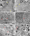
Transmission electron microscope (TEM) images of islet of Langerhans from saline/control (A, C, and E) and exenatide-treated pancreas (B, D, and F) (G. Finzi, S. La Rosa, and F. Folli, unpublished observations). Pancreatic specimens of four saline-treated and four exenatide-treated baboons were fixed in 2% paraformaldehyde and 2% glutaraldehyde (Karnovsky fixative), post-fixed in 1% osmium tetroxide, and embedded in Epon.Araldyte. Thin sections were counterstained with uranyl acetate and lead citrate and observed with a Philips/Morgagni/Thermo Fisher Scientific electron microscope (FEI Thermofisher Company, Eindhoven, the Netherlands). A and B: exenatide-treated pancreases show endocrine cells healthy and well granulated. C and D: TEM from control and exenatide-treated pancreas immunostained with anti-proinsulin antibodies. For ultrastructural immunocytochemistry, the sections were pretreated with sodium metaperiodate for 30 min, then placed onto a drop of 1% ovoalbumin for 5 min, transferred onto a drop of anti-proinsulin (monoclonal mouse anti-proinsulin DSHB, Gentofte, Denmark) diluted 1:10 overnight, then after rinses transferred onto a drop of 18 nm colloidal gold-AffiniPure (Jackson Immunoresearch, West Grove, PA) goat antimouse diluted 1:20 for 1 h, and, after rinses, counterstained. In controls experiments, primary antibodies were omitted. After exenatide treatment, β cells immature granules, showing a homogenous gray content and proinsulin labeled (red arrows), are less represented than in control pancreas. E and F: TEM from control and exenatide-treated animals immunostained with anti-proinsulin (18 nm colloidal gold) and anti-insulin (12 nm colloidal gold) antibodies. After sodium metaperiodate, thin sections were placed onto a drop of 1% ovoalbumin for 5 min, transferred onto a drop of anti-proinsulin (monoclonal mouse anti-proinsulin DSHB, Gentofte, Denmark) diluted 1:10 overnight, then after rinses transferred onto a drop of anti-insulin (polyclonal guinea pig anti-insulin, Dako, Glostrup, Denmark) diluted 1:50 overnight, after subsequent rinses transferred onto a mixture of 18 nm colloidal gold-AffiniPure (Jackson Immunoresearch, West Grove, PA) goat anti-mouse diluted 1:20 and of 12 nm colloidal gold-AffiniPure (Jackson Immunoresearch, West Grove, PA) donkey anti-guinea pig diluted 1:20, and finally counterstained after rinses. Controls experiments were done with primary antibodies omission. After saline treatment, β cells granules are predominant immature, showing homogenous gray matrix (red arrows) and containing proinsulin. After exenatide treatment β cells exhibit numerous mature granules, characterized by dense cores and peripheral clear halos (white arrows) and insulin content, beyond immature granules.
Figure 4.
Figure 4.
Amino acid sequence homology in glucagon-like peptide 1 (GLP-1, blue), glucose-dependent insulinotropic polypeptide (GIP, orange), and glucagon-like peptide-1 receptor (GLP-1R) agonists, and dual incretin agonists (gray). The DDP-4 cleavage site is reported in red.

References

    1. Samols E, Marri G, Marks V. Promotion of insulin secretion by glucagon. Lancet 2: 415–416, 1965. doi:10.1016/s0140-6736(65)90761-0. - DOI - PubMed
    1. Ma X, Zhang Y, Gromada J, Sewing S, Berggren PO, Buschard K, Salehi A, Vikman J, Rorsman P, Eliasson L. Glucagon stimulates exocytosis in mouse and rat pancreatic alpha-cells by binding to glucagon receptors. Mol Endocrinol 19: 198–212, 2005. doi:10.1210/me.2004-0059. - DOI - PubMed
    1. Capozzi ME, DiMarchi RD, Tschöp MH, Finan B, Campbell JE. Targeting the incretin/glucagon system with triagonists to treat diabetes. Endocr Rev 39: 719–738, 2018. doi:10.1210/er.2018-00117. - DOI - PMC - PubMed
    1. Tschöp MH, Finan B, Clemmensen C, Gelfanov V, Perez-Tilve D, Müller TD, DiMarchi RD. Unimolecular polypharmacy for treatment of diabetes and obesity. Cell Metab 24: 51–62, 2016. doi:10.1016/j.cmet.2016.06.021. - DOI - PubMed
    1. Labarre J. Sur les possibilities d’un traitement du diabete par. L’incretine. Bull Acad R Med Belg 12: 620–634, 1932.

Publication types

MeSH terms