. 2023 Sep 22;9(38):eadg0488.
doi: 10.1126/sciadv.adg0488. Epub 2023 Sep 20.

Single-cell genotypic and phenotypic analysis of measurable residual disease in acute myeloid leukemia

Affiliations

Single-cell genotypic and phenotypic analysis of measurable residual disease in acute myeloid leukemia

Troy M Robinson et al. Sci Adv. .

Abstract

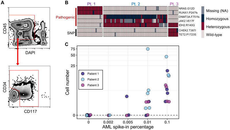
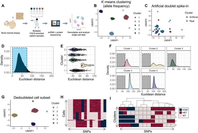
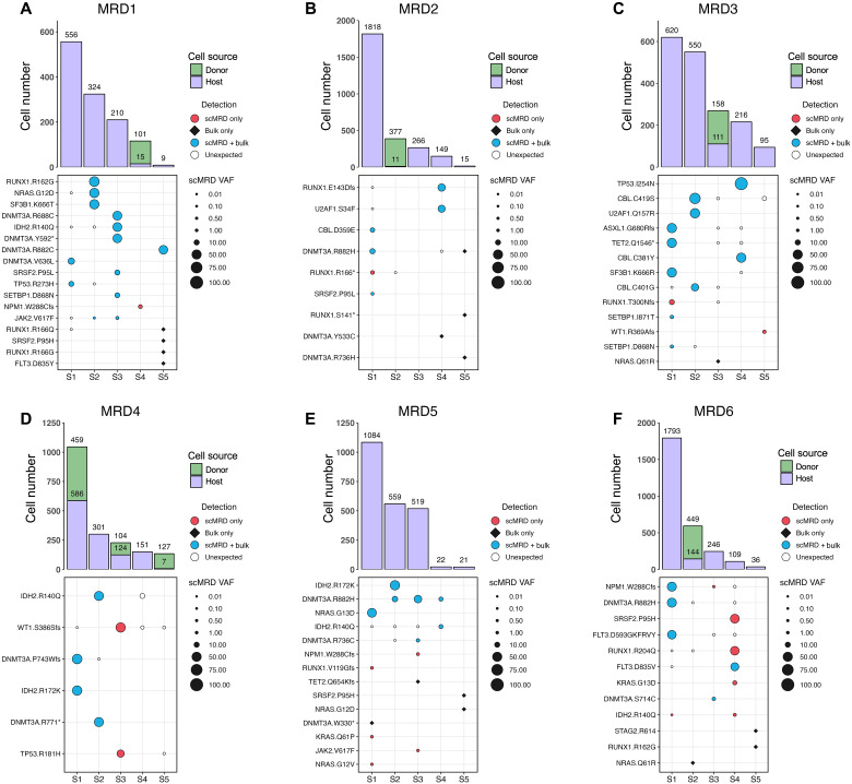
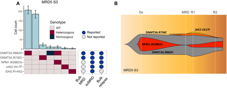
Measurable residual disease (MRD), defined as the population of cancer cells that persist following therapy, serves as the critical reservoir for disease relapse in acute myeloid leukemia and other malignancies. Understanding the biology enabling MRD clones to resist therapy is necessary to guide the development of more effective curative treatments. Discriminating between residual leukemic clones, preleukemic clones, and normal precursors remains a challenge with current MRD tools. Here, we developed a single-cell MRD (scMRD) assay by combining flow cytometric enrichment of the targeted precursor/blast population with integrated single-cell DNA sequencing and immunophenotyping. Our scMRD assay shows high sensitivity of approximately 0.01%, deconvolutes clonal architecture, and provides clone-specific immunophenotypic data. In summary, our scMRD assay enhances MRD detection and simultaneously illuminates the clonal architecture of clonal hematopoiesis/preleukemic and leukemic cells surviving acute myeloid leukemia therapy.


Figures

Fig. 1.
Fig. 1.. Limit of mutation detection with the scMRD assay.
(A) Schema of the gating strategy used for flow cytometric enrichment of live CD34+ and/or CD117+ cells after spiking AML cells into normal bone marrow. For clinical MRD samples included in this study, the abnormal blasts were confirmed to be positive for CD34 and/or CD117. (B) Representative heatmap showing mutation calling of spiked-in AML blasts in a limiting dilution experiment testing a sensitivity of 0.1%. Each column represents a single cell. (C) Summary of mutation detection at various sensitivity levels. This plot represents two independent experiments. NA, not applicable.
Fig. 2.
Fig. 2.. Workflow and computational demultiplexing of scMRD data.
(A) Schema of scMRD workflow (generated via BioRender). (B) Predoublet exclusion K-means clustering and UMAP analysis of SNP allele frequencies in cells perfectly genotyped for the top 14 SNPs. (C) UMAP plot showing the results of clustering real cells (blue) with artificial doublets (red). (D) Distribution of Euclidean distances from real cells to their respective cluster centers. (E) Violin plot showing by-cluster Euclidean distances of each real cell to the respective cluster center. (F) By-cluster Euclidean distances of each real cell to an artificial doublet cluster center. (G) Postdoublet exclusion K-means clustering and UMAP analysis of SNP allele frequencies. (H) Heatmap showing SNP genotypes in singlet clusters. (I) Heatmap showing the most common SNP profile for each cluster. (B) to (I) show a representative example (MRD5) of the computational pipeline output. UMAP, uniform manifold approximation and projection; HOM, homozygous; HET, heterozygous; WT, wild-type.
Fig. 3.
Fig. 3.. Deconvolution plots for individual scMRD runs.
(A to F) Computationally recovered cell number per sample (top) and VAF of mutations detected by scMRD, bulk NGS, or both assays (bottom). Each plot represents a single multiplexed run, and each column corresponds to a unique patient sample. “Unexpected” detection denotes mutations detected by scMRD but not reported at any bulk NGS time point (diagnosis, remission, and relapse). For MRD2, we did not detect mutations in two samples (MRD2-S3 and MRD2-S5) and, thus, could not confirm the identity of these samples. However, these samples had a considerable difference in cell input for sequencing and cell recovery after deconvolution (see table S5). We reasoned that the sample with higher cell number input corresponded to the sample with much higher recovery. MRD1-S4, MRD2-S2, MRD3-S3, MRD4-S1/S3/S5, and MRD6-S2 were post–allo-HSCT. MRD3-S3 and MRD6-S2 were from the same patient but obtained at different time points.
Fig. 4.
Fig. 4.. Clonal analysis of MRD5-S3.
(A) Clonal barplot of a patient (MRD5-S3) illustrating scMRD-specific detection of NPM1 and JAK2 mutations that were present at relapse. (B) Fish plot showing clonal evolution based on bulk NGS at diagnosis, remission (6 month after diagnosis), relapse time point 1 (R1; 7 months after diagnosis), and relapse time point 2 (R2; 11 months after diagnosis).
Fig. 5.
Fig. 5.. Clone- and mutation-specific immunophenotype.
(A) Clone-specific immunophenotype. (B) Differential surface marker expression between CH/preleukemic versus leukemic clones. (C and D) UMAP analysis of immunophenotypes of single-mutant versus compound-mutant clones. Distribution of cells in UMAP space with cells colored by genotype (C) and UMAP depiction of cell immunophenotype including markers for T cells (CD3), myeloid cells (CD11b and CD33), stem/progenitors (CD117 and CD34), monocytes (CD14 and CD64), granulocytes (CD16), B cells (CD19), and erythroid cells (CD71) (D). Data are centered log ratio–normalized, centered, and scaled on a by-run basis.
Fig. 6.
Fig. 6.. scDNA + protein analysis enables simultaneous identification of donor cells and MRD.
(A) Aggregated deconvolution plot showing mutations detected and host-donor chimerism of post–allo-HSCT samples from six different patients included in the study. MRD6-S2 and MRD3-S3 were from the same patients; therefore, only the former is shown. MRD2-S2 had no mutations detected by either bulk NGS or scMRD assay. MRD4-S3 had an HDAC1 p.P243L mutation not covered by the scMRD panel. Refer to Fig. 4 for multiplex context for each of these samples. (B) Row-scaled heatmap of differential surface maker expression between donor and host cells in MRD1-S4. (C) Concordance of immunophenotype of MRD cells between MFC and scMRD in MRD1-S4. Number of cells analyzed for (B) and (C): donor WT = 97, Host WT = 6, and Host NPM1-mut = 13.

References

    1. H. Döhner, A. H. Wei, F. R. Appelbaum, C. Craddock, C. D. DiNardo, H. Dombret, B. L. Ebert, P. Fenaux, L. A. Godley, R. P. Hasserjian, R. A. Larson, R. L. Levine, Y. Miyazaki, D. Niederwieser, G. Ossenkoppele, C. Röllig, J. Sierra, E. M. Stein, M. S. Tallman, H.-F. Tien, J. Wang, A. Wierzbowska, B. Löwenberg, Diagnosis and management of AML in adults: 2022 ELN recommendations from an international expert panel. Blood 140, 2022, 1345–1377 - PubMed
    1. M. Heuser, S. D. Freeman, G. J. Ossenkoppele, F. Buccisano, C. S. Hourigan, L. L. Ngai, J. M. Tettero, C. Bachas, C. Baer, M. C. Béné, V. Bücklein, A. Czyz, B. Denys, R. Dillon, M. Feuring-Buske, M. L. Guzman, T. Haferlach, L. Han, J. K. Herzig, J. L. Jorgensen, W. Kern, M. Y. Konopleva, F. Lacombe, M. Libura, A. Majchrzak, L. Maurillo, Y. Ofran, J. Philippe, A. Plesa, C. Preudhomme, F. Ravandi, C. Roumier, M. Subklewe, F. Thol, A. A. van de Loosdrecht, B. A. van der Reijden, A. Venditti, A. Wierzbowska, P. J. M. Valk, B. L. Wood, R. B. Walter, C. Thiede, K. Döhner, G. J. Roboz, J. Cloos, 2021 update on MRD in acute myeloid leukemia: A consensus document from the European LeukemiaNet MRD Working Party. Blood 138, 2753–2767 (2021). - PMC - PubMed
    1. M. Terwijn, W. L. J. van Putten, A. Kelder, V. H. J. van der Velden, R. A. Brooimans, T. Pabst, J. Maertens, N. Boeckx, G. E. de Greef, P. J. M. Valk, F. W. M. B. Preijers, P. C. Huijgens, A. M. Dräger, U. Schanz, M. Jongen-Lavrecic, B. J. Biemond, J. R. Passweg, M. van Gelder, P. Wijermans, C. Graux, M. Bargetzi, M. C. Legdeur, J. Kuball, O. de Weerdt, Y. Chalandon, U. Hess, L. F. Verdonck, J. W. Gratama, Y. J. M. Oussoren, W. J. Scholten, J. Slomp, A. N. Snel, M. C. Vekemans, B. Löwenberg, G. J. Ossenkoppele, G. J. Schuurhuis, High prognostic impact of flow cytometric minimal residual disease detection in acute myeloid leukemia: Data from the HOVON/SAKK AML 42A study. J. Clin. Oncol. 31, 3889–3897 (2013). - PubMed
    1. D. Araki, B. L. Wood, M. Othus, J. P. Radich, A. B. Halpern, Y. Zhou, M. Mielcarek, E. H. Estey, F. R. Appelbaum, R. B. Walter, Allogeneic hematopoietic cell transplantation for acute myeloid leukemia: Time to move toward a minimal residual disease-based definition of complete remission? J. Clin. Oncol. 34, 329–336 (2016). - PMC - PubMed
    1. C. S. Hourigan, L. W. Dillon, G. Gui, B. R. Logan, M. Fei, J. Ghannam, Y. Li, A. Licon, E. P. Alyea, A. Bashey, H. J. Deeg, S. M. Devine, H. F. Fernandez, S. Giralt, M. Hamadani, A. Howard, R. T. Maziarz, D. L. Porter, B. L. Scott, E. D. Warlick, M. C. Pasquini, M. E. Horwitz, Impact of conditioning intensity of allogeneic transplantation for acute myeloid leukemia with genomic evidence of residual disease. J. Clin. Oncol. 38, 1273–1283 (2020). - PMC - PubMed