. 2023 Oct 5;110(10):1769-1786.
doi: 10.1016/j.ajhg.2023.08.012. Epub 2023 Sep 19.

Systematically testing human HMBS missense variants to reveal mechanism and pathogenic variation

Affiliations

Systematically testing human HMBS missense variants to reveal mechanism and pathogenic variation

Warren van Loggerenberg et al. Am J Hum Genet. .

Abstract

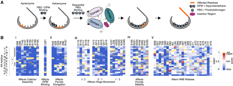
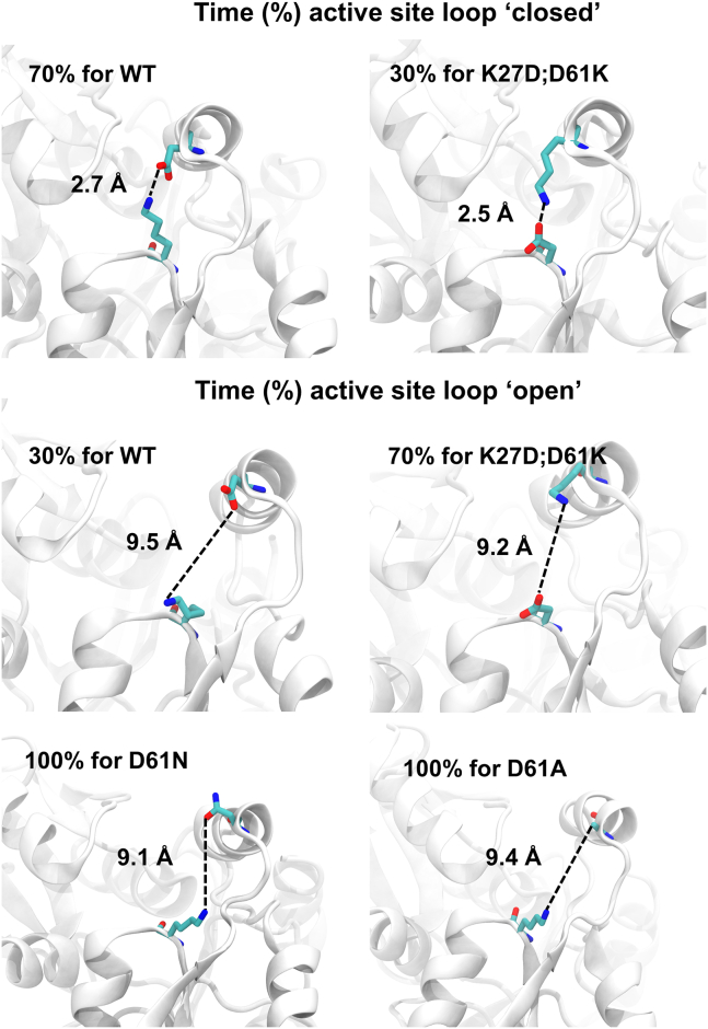
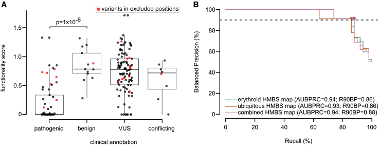
Defects in hydroxymethylbilane synthase (HMBS) can cause acute intermittent porphyria (AIP), an acute neurological disease. Although sequencing-based diagnosis can be definitive, ∼⅓ of clinical HMBS variants are missense variants, and most clinically reported HMBS missense variants are designated as "variants of uncertain significance" (VUSs). Using saturation mutagenesis, en masse selection, and sequencing, we applied a multiplexed validated assay to both the erythroid-specific and ubiquitous isoforms of HMBS, obtaining confident functional impact scores for >84% of all possible amino acid substitutions. The resulting variant effect maps generally agreed with biochemical expectations and provide further evidence that HMBS can function as a monomer. Additionally, the maps implicated specific residues as having roles in active site dynamics, which was further supported by molecular dynamics simulations. Most importantly, these maps can help discriminate pathogenic from benign HMBS variants, proactively providing evidence even for yet-to-be-observed clinical missense variants.

Keywords: AIP; HMBS,; acute hepatic porphryia; acute intermittent porphryia; clinical variant interpretation; deep mutational scanning; heme biosynthesis; hydroxymethylbilane synthase; molecular dynamics; variant effect mapping.


Conflict of interest statement

Declaration of interests F.P.R. is an investor in Ranomics, Inc., and is an investor in and advisor for SeqWell, Inc., BioSymetrics, Inc., and Constantiam Biosciences, Inc., and has accepted conference travel support from Illumina, Inc. L.F., A.C., and R.N. are employed by and invested in Invitae. R.J.D. has received both a grant and royalties and has also served as a consultant for Alnylam Pharmaceuticals.

Figures

Figure 1
Figure 1
Generating and evaluating HMBS variant effect maps (A) Workflow for generating HMBS variant effect maps. (B) Correspondence between erythroid-specific and ubiquitous HMBS isoform functional scores. For reference, null- and WT-like scores are indicated with dashed blue or gold lines, respectively, while the red line corresponds to a linear regression fit (R = 0.96; p = 2.2 × 10−16). (C) Distributions of functional impact scores of nonsense (blue), synonymous (gold), and missense variants (gray) from the combined erythroid-specific and ubiquitous HMBS map. (D) Preview of full-sized combined HMBS map.
Figure 2
Figure 2
Identifying patterns of mutational tolerance (A) An overview of pyrrole-binding sites and structural fluctuations of HMBS involved in polypyrrole elongation and HMB formation. (B) Functional scores for each possible substituted amino acid (y axis) at each active-site residue position (x axis) responsible for (I) altering cofactor binding, (II) PBG binding for pyrrole chain elongation, (III) hinge flexibility, (IV) pyrrole stability, and (V) HMB release. For each substitution, diagonal bar sizes convey estimated measurement error in the corresponding functional score. Box color either indicates the WT residue (yellow); a substitution with damaging (blue), tolerated (white), or above-WT (“hyper-complementing,” red) functional score; or missing data (gray).
Figure 3
Figure 3
Variant impact on “closed” and “open” active site loop conformations The average distance (Å) between protein position 27 and 61 is shown, along with fraction of time spent in each conformation, for WT HMBS and three HMBS variants—p.Asp61Asn, p.Asp61Ala, and the double mutant p.Asp61Lys;Lys27Asp.
Figure 4
Figure 4
Modeling the effects of HMBS missense variants on protein stability and structure (A) Comparison of functional impact scores (black) and predicted free energy change (ΔΔG; red) values of HMBS missense variants. Plotted values are averages within windows of five amino acid (AA) positions. (B) WT (top; I–IV) and p.Glu250Arg variant (bottom; V–VIII) comparison. The p.Glu250Arg substitution repels Arg116 and opens a channel (V), which is exposed to solvent (VI). The PBG-Gly218 interaction is lost in the p.Glu250Arg variant (VII) and replaced by a salt bridge between PBG and Arg195, which in turn disrupts Asp99-pyrrol interactions (VIII). For clarity, hydrogen atoms are not shown. Water molecules (represented as red spheres) that are within 7 Å of either Glu250/Arg250 or Gln217 for more than 50% of the simulation time are shown. (C) Structural model of HMBS; colored according to the median functionality score of substitutions at each position, along with a wireframe model of the tetrapyrrole (green), and noting residues located at the dimer interface. (D) Median functionality scores of variants at amino acid positions that were (1) below 20% accessible surface area (ASA), (2) above 40% ASA, (3) at the dimerization interface with a threshold ΔASA of 1.0, and (4) active site residues required for polypyrrole assembly. Boxes correspond to interquartile range, and bold bars indicate medians. Whiskers correspond to minima and maxima. p values were calculated by Mann-Whitney U test.
Figure 5
Figure 5
Performance of variant effect maps in distinguishing pathogenic from benign reference variants (A) The distribution of functional impact scores for reference “pathogenic,” “benign,” “VUS,” and “conflicting” variant sets. Variants from residue positions 160 to 215, 255, or 355 (indicated in red) were excluded from performance analysis given the suspected limitation of our assay for these variants. Boxes correspond to interquartile range, and bold bars indicate medians. Whiskers correspond to minima and maxima. Significance was evaluated with a Mann-Whitney U test. (B) Evaluation of precision (fraction of variants scoring below each threshold functionality score that are in the positive reference set containing pathogenic variants) vs. recall (fraction of positive reference variants with functionality scores below threshold). Here, precision values have been “balanced” to reflect performance in a setting where positive and negative sets contain the same number of variants. Balanced precision-recall curves are shown for erythroid-specific (green), ubiquitous (orange), and combined maps (pink). Performance is also described in terms of area under the balanced precision vs. recall curve (AUBPRC) and recall at a balanced precision of 90% (R90BP).

Update of

References

    1. Bissell D.M., Anderson K.E., Bonkovsky H.L. N. Engl. J. Med. 2017;377:862–872. - PubMed
    1. Chen B., Solis-Villa C., Hakenberg J., Qiao W., Srinivasan R.R., Yasuda M., Balwani M., Doheny D., Peter I., Chen R., Desnick R.J. Acute Intermittent Porphyria: Predicted Pathogenicity of HMBS Variants Indicates Extremely Low Penetrance of the Autosomal Dominant Disease. Hum. Mutat. 2016;37:1215–1222. - PMC - PubMed
    1. Baumann K., Kauppinen R. Penetrance and predictive value of genetic screening in acute porphyria. Mol. Genet. Metab. 2020;130:87–99. - PubMed
    1. Lenglet H., Schmitt C., Grange T., Manceau H., Karboul N., Bouchet-Crivat F., Robreau A.-M., Nicolas G., Lamoril J., Simonin S., et al. From a dominant to an oligogenic model of inheritance with environmental modifiers in acute intermittent porphyria. Hum. Mol. Genet. 2018;27:1164–1173. - PubMed
    1. Grandchamp B., De Verneuil H., Beaumont C., Chretien S., Walter O., Nordmann Y. Tissue-specific expression of porphobilinogen deaminase. Two isoenzymes from a single gene. Eur. J. Biochem. 1987;162:105–110. - PubMed

MeSH terms

Substances