Case Reports
. 2023 Sep 20;16(9):e256722.
doi: 10.1136/bcr-2023-256722.

Aortic sarcoma: a challenging diagnosis

Affiliations
Case Reports

Aortic sarcoma: a challenging diagnosis

Thi Huong Giang Bui et al. BMJ Case Rep. .

Abstract

Sarcomas of the aorta are rare tumours with various clinical presentations. The most common symptoms are embolic events, constitutional symptoms, claudication, abdominal complaints, aneurysm/pseudoaneurysm, back pain and hypertension. We present a case of a woman in her early 60s having fever, fatigue and cough for 3 months. The chest CT revealed an aneurysm measuring 64.1×65.6 mm. The oncology and thoracic surgical teams were consulted and decided to do an open repair of the aorta and take specimens for histopathological examination, which later confirmed a pleomorphic undifferentiated sarcoma of the aorta. She was temporarily discharged on day 9th after the surgery, followed up by chemotherapy in subsequent admission.

Keywords: Oncology; Surgical oncology; Vascular surgery.


Conflict of interest statement

Competing interests: None declared.

Figures

Figure 1
Figure 1
The CT scan revealed a descending aortic aneurysm 1 year prior.
Figure 2
Figure 2
The stent graft placement in the descending aorta at the level of aneurysm after the patient getting a diagnosis 1 year ago.
Figure 3
Figure 3
Preoperative chest CT revealed an aneurysm (with a stent graft placed 1 year ago) in the thoracic aorta, later confirmed by biopsy as a sarcoma.
Figure 4
Figure 4
Preoperative MRI revealed an aortic aneurysm with hyperintensity in T1-weighted images.
Figure 5
Figure 5
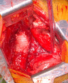
The mass was removed, and the aorta was reconstructed using an artificial graft.
Figure 6
Figure 6
H&E stain of the mass biopsy specimen showed diverse cell morphology with nuclear pleomorphism and numerous mitoses.
Figure 7
Figure 7
CD68 staining was positive.

References

    1. Rusthoven CG, Liu AK, Bui MM, et al. Sarcomas of the aorta: a systematic review and pooled analysis of published reports. Ann Vasc Surg 2014;28:515–25. 10.1016/j.avsg.2013.07.012 - DOI - PubMed
    1. Seddon B, Strauss SJ, Whelan J, et al. Gemcitabine and Docetaxel versus doxorubicin as first-line treatment in previously untreated advanced Unresectable or metastatic soft-tissue Sarcomas (Geddis): a randomised controlled phase 3 trial. Lancet Oncol 2017;18:1397–410. 10.1016/S1470-2045(17)30622-8 - DOI - PMC - PubMed
    1. Chiche L, Mongrédien B, Brocheriou I, et al. Primary tumors of the Thoracoabdominal aorta: surgical treatment of 5 patients and review of the literature. Ann Vasc Surg 2003;17:354–64. 10.1007/s10016-003-0018-x - DOI - PubMed
    1. Whittington EA, Duncan LD, McNally MM. Pleomorphic undifferentiated aortic sarcoma presenting as persistent Endoleak after Endovascular aneurysm repair. J Vasc Surg Cases Innov Tech 2019;5:294–7. 10.1016/j.jvscit.2019.04.006 - DOI - PMC - PubMed
    1. von Falck C, Meyer B, Fegbeutel C, et al. Imaging features of primary Sarcomas of the great vessels in CT, MRI and PET/CT: a single-center experience. BMC Med Imaging 2013;13:25. 10.1186/1471-2342-13-25 - DOI - PMC - PubMed

Publication types