. 2023 Sep 20;14(1):5832.
doi: 10.1038/s41467-023-41391-7.

NLRP3 selectively drives IL-1β secretion by Pseudomonas aeruginosa infected neutrophils and regulates corneal disease severity

Affiliations

NLRP3 selectively drives IL-1β secretion by Pseudomonas aeruginosa infected neutrophils and regulates corneal disease severity

Martin S Minns et al. Nat Commun. .

Abstract

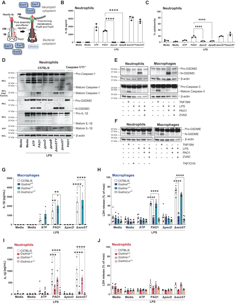
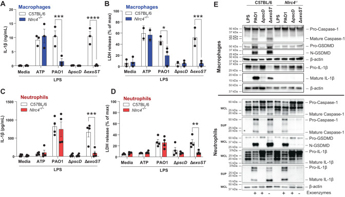
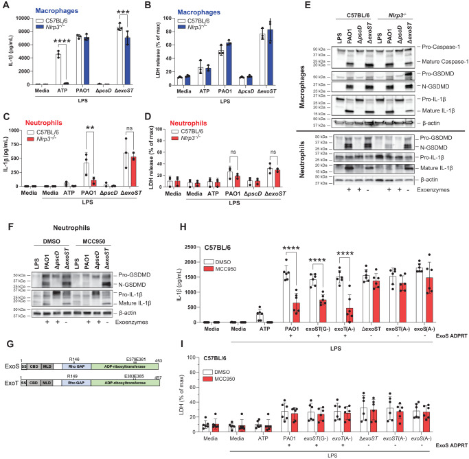
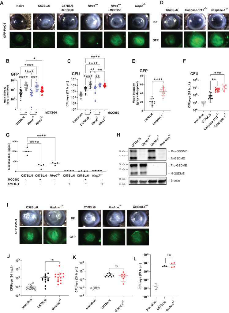
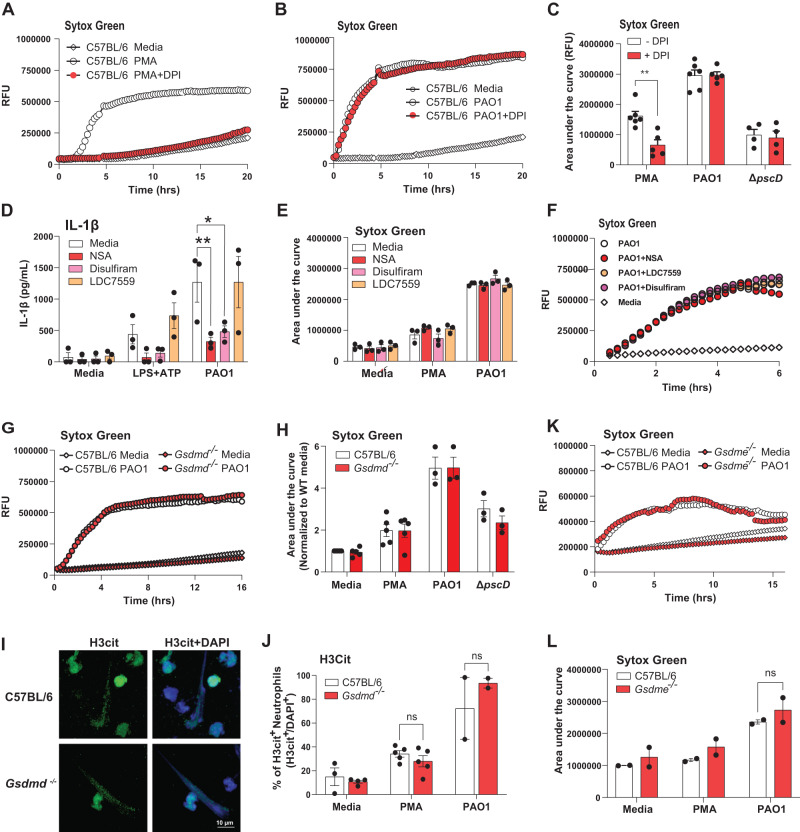
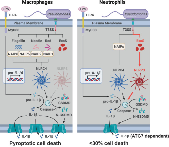
Macrophages infected with Gram-negative bacteria expressing Type III secretion system (T3SS) activate the NLRC4 inflammasome, resulting in Gasdermin D (GSDMD)-dependent, but GSDME independent IL-1β secretion and pyroptosis. Here we examine inflammasome signaling in neutrophils infected with Pseudomonas aeruginosa strain PAO1 that expresses the T3SS effectors ExoS and ExoT. IL-1β secretion by neutrophils requires the T3SS needle and translocon proteins and GSDMD. In macrophages, PAO1 and mutants lacking ExoS and ExoT (ΔexoST) require NLRC4 for IL-1β secretion. While IL-1β release from ΔexoST infected neutrophils is also NLRC4-dependent, infection with PAO1 is instead NLRP3-dependent and driven by the ADP ribosyl transferase activity of ExoS. Genetic and pharmacologic approaches using MCC950 reveal that NLRP3 is also essential for bacterial killing and disease severity in a murine model of P. aeruginosa corneal infection (keratitis). Overall, these findings reveal a function for ExoS ADPRT in regulating inflammasome subtype usage in neutrophils versus macrophages and an unexpected role for NLRP3 in P. aeruginosa keratitis.


Conflict of interest statement

The authors declare that they have no competing interests.

Figures

Fig. 1
Fig. 1. The role of T3SS, GSDMD and GSDME in IL-1β secretion by P. aeruginosa infected neutrophils.
A P. aeruginosa T3SS is preassembled and spans the bacterial cell envelope (IM, inner membrane, OM, outer membrane and PG, peptidoglycan layer). Following cell contact, the pore-forming translocator proteins, PopB and PopD, assemble into a pore, which docks to the needle tip (PcrV, red). Effector secretion is triggered, resulting in export of effector proteins across the bacterial cell envelope and host cell plasma membrane (PM) into the cytosol of the targeted cell. P. aeruginosa strain PAO1 used in the current study expresses ExoS, ExoT, and ExoY, but not ExoU. B Quantification of IL-1β by ELISA, and C Cell death quantified by lactose dehydrogenase (LDH) activity in the supernatant of infected cells. D Caspase-1, GSDMD and IL-1β cleavage in whole cell lysates (WCL) and supernatants (SUP) from C57BL/6 and caspase-1/11-/- bone marrow neutrophils primed 3 h with LPS and incubated 1 h with ATP or 1 h with P. aeruginosa strain PAO1 or PAO1 mutants ∆pscD (no needle), ∆popB (no translocon), ∆exoSTY or ∆exoST. E, F Caspase-3, GSDMD and GSDME cleavage in lysates with supernatants from C57BL/6 bone marrow neutrophils and bone marrow derived macrophages primed with LPS and infected with PAO1 in the presence of ZVAD. TNF-α and SMAC mimetic (TNF/SM) induced caspase-3 / GSDME cleavage after 4 h (no LPS priming). GJ IL-1β secretion and LDH release from infected bone marrow-derived macrophages and bone marrow neutrophils from C57BL/6, Gsdmd-/- Gsdme -/- and Gsdmd/e-/- mice primed with LPS and incubated with PAO1 or indicated mutants. Western blots are representative of 3 repeat experiments. Each data point represents one independent experiment (3-4 biological replicates); error bars are mean + /- standard deviation (SD). Statistical significance was assessed by 1-way ANOVA followed by a Brown-Forsythe post-test (B, C), or using 2-way ANOVA followed by Tukey’s post-test (G-J). For all figures, ****p < 0.0001, ***p < 0.001; **<0.01.
Fig. 2
Fig. 2. NLRC4 independent IL-1β secretion by PAO1 infected neutrophils.
Bone marrow derived macrophages (A, B) and bone marrow neutrophils (C, D) from C57BL/6 and Nlrc4-/- mice were primed 3 h with LPS and infected with PAO1, ∆pscD (no needle), ∆popB (no translocon), or ∆exoST. Quantification of IL-1β secretion by ELISA (A, C) and LDH release (B, D). E Caspase-1, GSDMD and IL-1β cleavage in infected macrophages and neutrophils from C57BL/6 and Nlrc4-/- mice were examined by western blot. Macrophage westerns are combined whole cell lysate and TCA precipitated supernatants. Neutrophil whole cell lysates (WCL) and supernatants (SUP) were examined separately. Western blots are representative of 3 repeat experiments. Each data point in panels A-D represents one independent experiment (3-4 biological replicates). Statistical significance was assessed by 2-way ANOVA followed by Tukey’s post-test. Error bars are mean +/- SD.
Fig. 3
Fig. 3. PAO1 mediated IL-1β secretion by neutrophils is NLRP3 dependent in the presence of ExoS ADPRT.
Bone marrow derived macrophages (A, B) and bone marrow neutrophils (C, D) from C57BL/6 and Nlrp3-/- mice were incubated with PAO1, ∆pscD or ∆exoST. IL-1β secretion and LDH release were quantified. E GSDMD and IL-1β cleavage was examined by western blot in macrophages and neutrophils from Nlrp3-/- mice. F GSDMD and IL-1β cleavage in C57BL/6 neutrophils infected with PAO1 or mutants in the presence of the NLRP3 inhibitor MCC950. Protein cleavage was examined in lysates combined with TCA precipitated supernatants. G GTPase (GAP) and ADP ribosyltransferase (ADPRT) regions of ExoS and ExoT showing amino acids required for enzymatic function and which are also the sites of point mutations. H, I Neutrophils from C57BL/6 mice infected with PAO1, ∆exoST or mutants with indicated point mutations (ExoS, ExoT A-, G-) in the presence of MCC950. Strains expressing ExoS ADPRT are indicated. Western blots are representative of 3 repeat experiments. For panels AD, H, I, each data point represents one independent experiments (3-6 biological repeats). Error bars are mean +/- SD. Statistical significance was assessed by 2-way ANOVA followed by Tukey’s post-test.
Fig. 4
Fig. 4. The role of inflammasomes, caspase-1, GSDMD and GSDME in P. aeruginosa corneal infections.
A-C Corneas of MCC950 treated C57BL/6, Nlrc4-/-, and Nlrp3-/- mice were infected with 5×10⁴ GFP expressing PAO1. After 24 h, corneal opacification and total GFP bacteria were quantified by image analysis, and viable bacteria were measured by CFU. A representative images of infected corneas and GFP-PAO1. B Quantification of GFP in infected corneas. C CFU in infected corneas. DF Corneal opacification, GFP-PAO1 and CFU in C57BL/6, caspase-1 and caspase-1/11-/- mice. G Bioactive IL-1β in infected corneas from C57BL/6, MCC950 treated mice, and Nlrp3-/- mice using the IL-1R reporter cells in the presence of neutralizing anti-IL-1β HL PAO1 infected C57BL/6, gsdmd-/-, gsdme -/- and gsdmd / gsdme -/- corneas. H GSDMD and GSDME cleavage in infected corneas. I Representative corneas, and CFU (J–L) GSDMD and GSDME cleavage products were examined by western blot. Western blots are representative of 3 repeat experiments. Each data point represents a single infected cornea from 3 independent experiments with 4 infected corneas. Statistical significance was assessed by 1-way ANOVA followed by Kruskal-Wallis post-test for in vivo analysis. **** represents p < 0.0001, *** is p < 0.001; ** is <0.01, * is <0.05.
Fig. 5
Fig. 5. PAO1 induced NETosis is independent of ROS, GSDMD and GSDME.
A–C Role of ROS in PMA stimulated, but not PAO1 infected peritoneal neutrophils. Representative time courses (A, B) and combined data from biological replicates (C). D-F IL-1β secretion (D) and NETosis (E) by peritoneal neutrophils incubated with reported GSDMD inhibitors necrosulfonamide (NSA), disulfiram, and LDC7559. NETosis was quantified by Sytox detection of extracellular DNA after 16 h (Area under the curve). Panel F is a representative time course. GJ Peritoneal neutrophils from C57BL/6 and Gsdmd-/- mice incubated 16 h with PMA, PAO1 or ∆pscD and extracellular DNA measured by Sytox. G representative time course, and H quantification of the area under the curve. I Representative images of citrullinated histone 3 (H3Cit) as an indicator of NETosis in PAO1 infected neutrophils (original and other images are in Fig. S4F). J Quantification of H3Cit+ neutrophils per high power field. K, L Representative time course and quantification of NETosis in C57BL/6 and Gsdme-/- mice. ** is <0.01, * is <0.05. Each data point represents one independent experiment (2-6 biological repeats). Error bars are mean +/- SEM. Statistical significance was assessed by 2-way ANOVA followed by Tukey’s post-test.
Fig. 6
Fig. 6. Predicted sequence of events in P. aeruginosa induced IL-1β secretion by macrophages and neutrophils.
See text for description. Created with BioRender.com.

References

    1. Broz P, Pelegrin P, Shao F. The gasdermins, a protein family executing cell death and inflammation. Nat. Rev. Immunol. 2020;20:143–157. doi: 10.1038/s41577-019-0228-2. - DOI - PubMed
    1. Liu X, Xia S, Zhang Z, Wu H, Lieberman J. Channelling inflammation: gasdermins in physiology and disease. Nat. Rev. Drug Discov. 2021;20:384–405. doi: 10.1038/s41573-021-00154-z. - DOI - PMC - PubMed
    1. Fu, J. & Wu, H. Structural mechanisms of NLRP3 inflammasome assembly and activation. Annu. Rev. Immunol. 41, 301–316 (2023). - PMC - PubMed
    1. Karmakar M, et al. Neutrophil IL-1beta processing induced by pneumolysin is mediated by the NLRP3/ASC inflammasome and caspase-1 activation and is dependent on K+ efflux. J. Immunol. 2015;194:1763–1775. doi: 10.4049/jimmunol.1401624. - DOI - PMC - PubMed
    1. Karmakar M, Katsnelson MA, Dubyak GR, Pearlman E. Neutrophil P2X7 receptors mediate NLRP3 inflammasome-dependent IL-1beta secretion in response to ATP. Nat. Commun. 2016;7:10555. doi: 10.1038/ncomms10555. - DOI - PMC - PubMed

Publication types

Substances