. 2023 Sep 20;9(1):348.
doi: 10.1038/s41420-023-01642-4.

Cdk12 maintains the integrity of adult axons by suppressing actin remodeling

Affiliations

Cdk12 maintains the integrity of adult axons by suppressing actin remodeling

L N Townsend et al. Cell Death Discov. .

Abstract

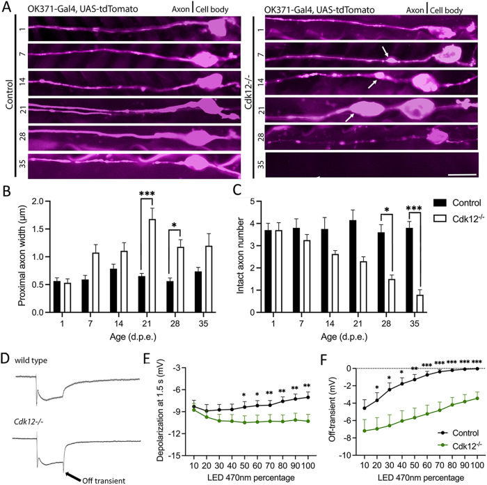
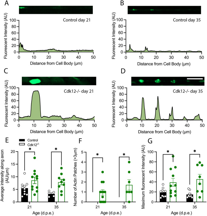
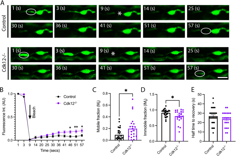
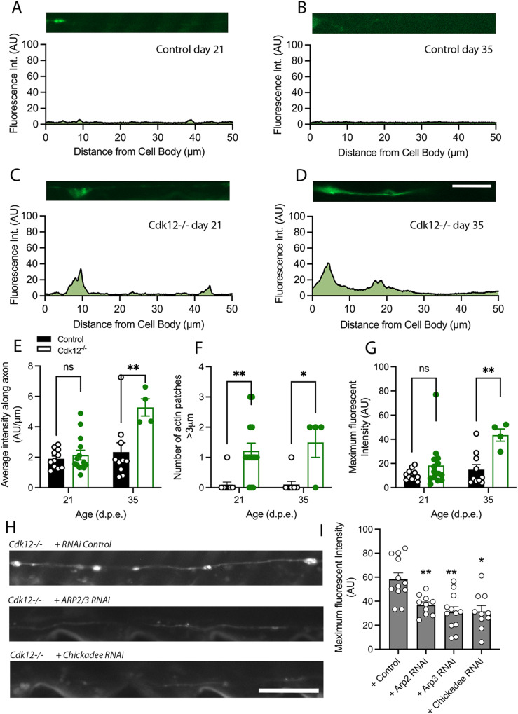
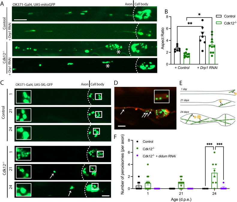
The role of cyclin-dependent kinases (CDKs) that are ubiquitously expressed in the adult nervous system remains unclear. Cdk12 is enriched in terminally differentiated neurons where its conical role in the cell cycle progression is redundant. We find that in adult neurons Cdk12 acts a negative regulator of actin formation, mitochondrial dynamics and neuronal physiology. Cdk12 maintains the size of the axon at sites proximal to the cell body through the transcription of homeostatic enzymes in the 1-carbon by folate pathway which utilize the amino acid homocysteine. Loss of Cdk12 leads to elevated homocysteine and in turn leads to uncontrolled F-actin formation and axonal swelling. Actin remodeling further induces Drp1-dependent fission of mitochondria and the breakdown of axon-soma filtration barrier allowing soma restricted cargos to enter the axon. We demonstrate that Cdk12 is also an essential gene for long-term neuronal survival and loss of this gene causes age-dependent neurodegeneration. Hyperhomocysteinemia, actin changes, and mitochondrial fragmentation are associated with several neurodegenerative conditions such as Alzheimer's disease and we provide a candidate molecular pathway to link together such pathological events.


Conflict of interest statement

The authors declare no competing interests.

Figures

Fig. 1
Fig. 1. Cdk12 limits axon width specifically in the region closest to the cell body.
A Mutations in the gene Cdk12 caused an age dependent increase in proximal axon width and the formation of axonal swellings in wing sensory neurons. B Cdk12/− axon width was increased 2–3-fold at both 21 and dpe. C Cdk12−/− axons began to degenerate at 28 days and a 75% loss of axons was recorded at 35 days p.e. D Example electroretinogram traces of wild type and Cdk12−/− animals at 100% light intensity. E Loss of Cdk12 was associated with increased depolarization in response to 470 nm light. F Loss of Cdk12 caused a significantly greater off-transient response compared to control. Data was analyzed by two-way ANOVA and significant differences annotated as p < 0.05*, p < 0.01**, & p < 0.001*** between genotypes. White arrows indicate axonal swellings. Graphs are expressed as Mean ± SEM and N = ≥10 wings for each group. Scale bars = 10 µm.
Fig. 2
Fig. 2. Cdk12 limits axonal b-actin patches.
A A representative b-actin fluorescence intensity plot of small focal b-actin patches in wild type in wing sensory neurons at 21 days. B Aged wild-type axons at 35 days also display small patches of actin. C Cdk12−/− axons display large axon patches that are intensely bright at 21 days. D By 35 days Cdk12−/− axons the b-actin fluorescence intensity plot displays multiple bright axon b-actin patches up to 50 μm away from cell body. E Cdk12−/− axons displayed a significantly greater area under the curve of fluorescent b-actin intensity plots. F Quantification shows that loss of Cdk12 is associated with increased b-actin patch formation. G The total fluorescence intensity of Cdk12−/− axons was greater than control at both 21 days and 35 days. Fluorescence was normalized to td-Tomato expressed in the same cell to label membranes. Data was analyzed by two-way ANOVA and significant differences annotated as p < 0.05* & p < 0.01** between genotypes. Graphs are expressed as Mean ± SEM and N = ≥ 8 wings for each group. Scale bar = 10 µm.
Fig. 3
Fig. 3. Cdk12 controls b-actin motility in the proximal axon.
A FRAP experiments at 21 days in wing sensory neurons show that b-actin fluorescence at the region of the axon proximal to the cell body shows minimal recovery at 57 s post bleach, whereas recovery was observed in Cdk12−/− axons. B maximum fluorescence recovery and plateau was seen at 46 s in all axons and a greater recovery was observed in Cdk12−/− axons. C Cdk12−/− axons display larger fraction of mobile b-actin compared to control. D Cdk12−/− axons have less immobile b-actin compared to control. E No difference in the time to recovery was observed between genotypes. Data was analyzed by two-way ANOVA or T-test and significant differences annotated as p < 0.05* & p < 0.01** between genotypes. The region of interest for FRAP experiments are indicated by the white circle. Graphs are expressed as Mean ± SEM and N = ≥ 20 wings for each group. Scale bar = 5 µm.
Fig. 4
Fig. 4. Cdk12 limits F-actin formation in axons.
A A representative F-actin fluorescence intensity plot of F-actin localization in wild type in wing sensory neurons at 21 days using genetically encoded LifeAct. F-actin is highly expressed in small patches closer to the cell body. B Aged wild-type axons at 35 days also display F-actin in proximal axon regions. C Cdk12−/− axons display large an increase in F-actin intensity at 21 days compared to age-matched controls. D By 35 days remaining Cdk12−/− axons display an F-actin fluorescence intensity plot that shows multiple bright axon F-actin patches up to 30 μm away from cell body. E Cdk12−/− axons displayed a significantly greater area under the curve of fluorescent F-actin intensity across 50 μm of axon. F Quantification shows that loss of Cdk12 is associated with increased F-actin patch formation. G The total fluorescence intensity of Cdk12−/− axons was significantly greater than control at 35 days. Fluorescence was normalized to td-Tomato expressed in the same cell to label membranes. H Chickadee and the Arp2/3 complex were required for F-actin formation I RNAis targeted against actin-binding proteins rescued the heightened actin phenotype. Data was analyzed by either one or two-way ANOVA and significant differences annotated as p < 0.05* & p < 0.01** between genotypes. Graphs are expressed as Mean ± SEM and N = ≥ 8 per group. Scale bars = 10 µm.
Fig. 5
Fig. 5. Cdk12 controls mitochondrial morphology and peroxisome position.
A Mitochondrial morphology was observed at 21 days in wing sensory neuronal clones. Cdk12−/− neurons and axonal swellings contain more spherical mitochondria compared to control, which was corrected with RNAi-mediated knockdown of Drp1 in axons. B Quantification shows that knockdown of the mitochondrial fission factor Drp1 caused an increase in mitochondrial aspect ratio in both wild-type and Cdk12 ablated axons. C Peroxisomes are largely confined to somato-dendritic regions in wild-type neurons at 1, 21, and 24 days, yet knock out of Cdk12 permitted age-dependent peroxisome entry into the proximal axon. D Peroxisomes in Cdk12−/− axons were present in axonal swellings and non-swollen regions at 21 days. E Illustration to show that peroxisomes may be permitted to enter Cdk12 ablated and aged axons on actin filaments via attachment to myosin motor proteins. F Quantification shows that at 24 days there are significantly more peroxisomes present in Cdk12−/− axonal clones compared to control, which can be rescued via RNAi-mediated knockdown of didum (Myosin V). Data was analyzed by two-way ANOVA and significant differences annotated as p < 0.05*, p < 0.01**, & p < 0.001*** between genotypes. Dashed lines define the soma-axon boundary, * define axonal swelling regions and arrows highlight axon localized peroxisomes. Graphs are expressed as Mean ± SEM and N = ≥ 8 wings for each group. Scale bars = 5 µm.
Fig. 6
Fig. 6. Cdk12 controls transcription of genes in the one-carbon by folate pathway, limiting homocysteine levels to allow for actin remodeling.
A A volcano plot showing differentially expressed genes from head tissue of Cdk12-/+ animals compared to wild-type controls. Pathway analysis revealed an enrichment of genes in the one-carbon pool by folate pathway. B A schematic to illustrate that transcriptional downregulation of genes in the one-carbon pool by folate ultimately lead to elevated homocysteine levels. C Quantification shows that brains of Cdk12-/+ animals have elevated homocysteine (Hcy) levels at 21 days compared to age-matched wild-types. D Addition of Hcy to human iPSC-derived cortical neurons revealed significant changes in F-actin morphology. E Actin is more discontinuous in the projections with Hcy treatment. F Actin was enriched in the cell bodies with Hcy treatment. RNA sequencing data is expressed as log2 foldchange and significance found in a DESeq2 analysis with an FDR adjusted P threshold of 0.05, indicated by the dashed lines. T-tests were used to analyze levels of Hcy in Drosophila brains and actin changes associated with Hcy treatment. Graphs are annotated as p < 0.001***. Graphs are expressed as Mean ± SEM, N = 4 (N = 8 heads per replicate) for RNA sequencing experiments. 3 replicates were used for iPSC experiment, N = 15 images for quantification. Scale bars = 50 µm and inset = 5 µm.

Similar articles

Cited by

References

    1. Grison A, Gaiser C, Bieder A, Baranek C, Atanasoski S. Ablation of cdk4 and cdk6 affects proliferation of basal progenitor cells in the developing dorsal and ventral forebrain. Dev Neurobiol. 2018;78:660–70. doi: 10.1002/dneu.22588. - DOI - PubMed
    1. Lange C, Huttner WB, Calegari F. Cdk4/cyclinD1 overexpression in neural stem cells shortens G1, delays neurogenesis, and promotes the generation and expansion of basal progenitors. Cell Stem Cell. 2009;5:320–31. doi: 10.1016/j.stem.2009.05.026. - DOI - PubMed
    1. Chen H-R, Juan H-C, Wong Y-H, Tsai J-W, Fann M-J. Cdk12 regulates neurogenesis and late-arising neuronal migration in the developing cerebral cortex. Cereb Cortex. 2017;27:2289–302. doi: 10.1093/cercor/bhw081. - DOI - PubMed
    1. Chen H-R, Lin G-T, Huang C-K, Fann M-J. Cdk12 and Cdk13 regulate axonal elongation through a common signaling pathway that modulates Cdk5 expression. Exp Neurol. 2014;261:10–21. doi: 10.1016/j.expneurol.2014.06.024. - DOI - PubMed
    1. Connell-Crowley L, Le Gall M, Vo DJ, Giniger E. The cyclin-dependent kinase Cdk5 controls multiple aspects of axon patterning in vivo. Curr Biol CB. 2000;10:599–602. doi: 10.1016/s0960-9822(00)00487-5. - DOI - PubMed