. 2023 Sep;621(7980):782-787.
doi: 10.1038/s41586-023-06538-y. Epub 2023 Sep 20.

The oldest three-dimensionally preserved vertebrate neurocranium

Affiliations

The oldest three-dimensionally preserved vertebrate neurocranium

Richard P Dearden et al. Nature. 2023 Sep.

Abstract

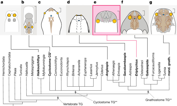
The neurocranium is an integral part of the vertebrate head, itself a major evolutionary innovation1,2. However, its early history remains poorly understood, with great dissimilarity in form between the two living vertebrate groups: gnathostomes (jawed vertebrates) and cyclostomes (hagfishes and lampreys)2,3. The 100 Myr gap separating the Cambrian appearance of vertebrates4-6 from the earliest three-dimensionally preserved vertebrate neurocrania7 further obscures the origins of modern states. Here we use computed tomography to describe the cranial anatomy of an Ordovician stem-group gnathostome: Eriptychius americanus from the Harding Sandstone of Colorado, USA8. A fossilized head of Eriptychius preserves a symmetrical set of cartilages that we interpret as the preorbital neurocranium, enclosing the fronts of laterally placed orbits, terminally located mouth, olfactory bulbs and pineal organ. This suggests that, in the earliest gnathostomes, the neurocranium filled out the space between the dermal skeleton and brain, like in galeaspids, osteostracans and placoderms and unlike in cyclostomes2. However, these cartilages are not fused into a single neurocranial unit, suggesting that this is a derived gnathostome trait. Eriptychius fills a major temporal and phylogenetic gap in our understanding of the evolution of the gnathostome head, revealing a neurocranium with an anatomy unlike that of any previously described vertebrate.


Conflict of interest statement

The authors declare no competing interests.

Figures

Fig. 1
Fig. 1. Overview of Eriptychius americanus PF 1795.
a,b, Photographs of part PF 1795a, which had the split face set in epoxy and was manually prepared (a), and its counterpart PF 1795b, which remains in rock matrix (b). Both are shown in an anatomically ventral view. c,d, Digital model of computed tomographic data of the combined part and counterpart with most of dermal skeleton rendered transparent: anatomical ventral view (corresponding to the visible area of the part in epoxy) (c) and anatomical dorsal view (buried in matrix in the counterpart) (d). Colour scheme for renders: blue-greys, cranial cartilages (matching the detailed scheme in Fig. 2); transparencies, the dermal skeleton; orange, branchial plates; red, orbital plates. Anterior to top in ad. ant. tess., anterior tesserae; artic. vent. tess., articulated ventral tesserae; branch. plate, branchial plate; cran. cart., cranial cartilages; disp., displaced; frag., fragment; L., left; orb. cart., orbital cartilage; orb. plates, orbital plates; R., right; vasc., vasculature; ?, probable. Scale bar applies to all panels.
Fig. 2
Fig. 2. The neurocranial cartilages of E. americanus PF 1795 based on computed tomography data.
ac, Cranial cartilages in estimated life position, with cartilages coloured in pairs in dorsal (a), ventral (b) and anterior (c) view. d,e, Mediolateral cartilages A in dorsal view (d) and median dorsal cartilage in ventral view (e) rendered with a vertical height map texture to emphasize the surface topology. f, Reconstruction of the forebrain relative to the cranial cartilages using a lamprey as a model,, shown in dorsal view. g, Cartilages in dorsal view, rendered transparent to show internal vasculature (red). h,i, Cartilages in preserved position in anterior view with dermal skeleton shown (h) and removed (i). Colours in a,b,c,f,h,i as in Fig. 1 with the following additions. Green, dermal skeleton. Red dashed line represents inferred position of mouth in c,h,i. In d and e lighter colours denote areas closer to the camera. Abbreviations as in Fig. 1 with the following additions: antorb. proc, antorbital process; ext. vasc. op., external vascular openings; forebr., forebrain; lat., lateral; medlat. cart, mediolateral cartilage; med. dors. cart, median dorsal cartilage; med., medial; med. vent. cart., median ventral cartilage; med. vent. ridge, median ventral ridge; olf. bulb, olfactory bulb; pin., pineal organ; pin. op., pineal opening; vent., ventral. Scale bar in a is shared by b,c; scale bar in d is shared by e.
Fig. 3
Fig. 3. The neurocrania of vertebrates in a phylogenetic framework.
a, Haikouichthys, adapted from ref. , CC-BY 4.0. b, Petromyzon. c, Generalized cyathaspid, adapted from ref. , Springer Nature. d, Sacabambaspis, reproduced from ref. , copyright ©1993 Elsevier Masson SAS, all rights reserved (but see ref. for alternative interpretation). e, Eriptychius. f, Shuyu, adapted from ref. , Springer Nature. g, Dicksonosteus adapted with permission from ref. ,Muséum national d’histoire naturelle, Paris. Phylogenetic scheme is the Adams consensus tree described in the text, bold taxon names represent those depicted above. Coloured regions illustrate positions of key sensory organs: yellow, eyes; blue, pineal. Light grey lines represent body outlines, black lines represent dermal body armour, light grey regions with dark grey borders represent cranial mineralizations, brown regions represent the brain (b), imprints of the brain in the dorsal headshield (c) or the endocast (f,g). Square brackets represent the preorbital part of the head. Petromyzon redrawn from Sketchfab (https://sketchfab.com/3d-models/lamprey-inside-c9f05e3f00c94d929e7ed018fac6d782) under a CC BY 4.0 licence. Abbreviations: CG, crown group; mand. gnath., mandibulate gnathostomes; TG, total group. *‘Crown-group cyclostomes’ represents the Petromyzontidae and Myxinoidea total groups and Gilpichthys, which was recovered in a polytomy with those two groups. **Cyclostome and gnathostome total groups in this topology recovered in a polytomy with Metaspriggina and (Haikouichthys + Myllokunmingia).
Extended Data Fig. 1
Extended Data Fig. 1. Eriptychius PF 1795 shown in the context of the surrounding matrix.
a,b, Tomograms showing sections through the part of the specimen preserved in the matrix. c–e, 3D render of the articulated Eriptychius specimen part preserved in epoxy relative to the matrix part, (c) in anatomical ventral view, (d) lateroventral view and (e) laterally with the matrix rendered transparent.
Extended Data Fig. 2
Extended Data Fig. 2. Photographs of Eriptychius PF 1795.
a, part preserved in matrix contrasting our interpretation with that of Denison. b, a close up of the front part of the specimen showing the vasculature. c, the part of the specimen preserved in epoxy with d, a close up of the ‘rostral plates and e, a close up of the orbital plates. Roman case labels, our interpretation; italics case labels, Denison’s interpretation corresponding to figure 2 of ref..
Extended Data Fig. 3
Extended Data Fig. 3. Eriptychius PF 1795 tissues in tomographic section.
Section of tomogram from the higher resolution scan set in coronal plane showing dermal ‘rostral’ plates overlying and wrapping cartilage.
Extended Data Fig. 4
Extended Data Fig. 4. 3D render of Eriptychius PF 1795.
a, in ventral view. b, in dorsal view. c, chain of orbital plates in visceral view, showing curved visceral surfaces. d,e, the best-visualized orbital plate in (d) lateral and (e) visceral view. f, g, left branchial plate in (f) lateral and (g) anterior view. h,?R. branchial plate fragment in lateral view. i, right branchial plate in lateral view. Colour scheme and abbreviations as in Figs. 1 and 2, except: grey, cartilages; blue-green, dermal skeleton. Lighter shades denote material from PF 1795a, darker shades PF 1795b.
Extended Data Fig. 5
Extended Data Fig. 5. 3D renders of the individual endocranial elements of Eriptychius PF 1795.
a, median dorsal cartilage in anterior view; b-d median ventral cartilage in (b) posterior, (c) left lateral and (d) dorsal view; e,f mediolateral cartilages A in (e) ventral and (f), posterior view; g,h both surfaces of mediolateral cartilages B; i,j both surfaces of mediolateral cartilages C; k-m, left orbital cartilage in (k) medial, (l) posterior and (m) latero-posterior view. Colour scheme and abbreviations as in Fig. 2.
Extended Data Fig. 6
Extended Data Fig. 6. Additional 3D renders of the vascularization of the endocranium of Eriptychius PF 1795.
a,b, median dorsal cartilage in (a) dorsal and (b) ventral view; c,d, median ventral cartilage in (c) ventral and (d) dorsal view; e,f, mediolateral cartilages A in (e) dorsal and (f) ventral view; g, mediolateral cartilages B; h, mediolateral cartilages C; i,j left orbital cartilage in (i) dorsal and (j) ventral view; k,l right orbital cartilage in (k) dorsal and (l) ventral view. Colour scheme as in Fig. 2.
Extended Data Fig. 7
Extended Data Fig. 7. Strict consensus result of the parsimony analysis.
Strict consensus tree resulting from the parsimony phylogenetic analysis described in the methods.
Extended Data Fig. 8
Extended Data Fig. 8. Adams consensus result of the parsimony analysis.
Adams consensus tree resulting from the parsimony phylogenetic analysis described in the methods.
Extended Data Fig. 9
Extended Data Fig. 9. Consensus result of the Bayesian analysis.
Majority rule consensus tree resulting from the Bayesian phylogenetic analysis described in the methods. Node values correspond to posterior probabilities.

Comment in

References

    1. Gans, C. in The Skull Vol. 2 (eds. Hanken, J. & Hall, B. K.) 1–35 (Chicago Univ. Press, 1993).
    1. Janvier, P. Early Vertebrates (Clarendon, 1996).
    1. Janvier, P. in The Skull Vol. 2 (eds. Hanken, J. & Hall, B. K.) 131–188 (Chicago Univ. Press, 1993).
    1. Shu D-G, et al. Head and backbone of the Early Cambrian vertebrate Haikouichthys. Nature. 2003;421:775–778. doi: 10.1038/nature01264. - DOI - PubMed
    1. Morris SC, Caron J. A primitive fish from the Cambrian of North America. Nature. 2014;512:419–422. doi: 10.1038/nature13414. - DOI - PubMed

Publication types