. 2023 Oct;42(44):3252-3259.
doi: 10.1038/s41388-023-02835-y. Epub 2023 Sep 21.

Neoadjuvant botensilimab plus balstilimab response pattern in locally advanced mismatch repair proficient colorectal cancer

Affiliations

Neoadjuvant botensilimab plus balstilimab response pattern in locally advanced mismatch repair proficient colorectal cancer

Pashtoon Murtaza Kasi et al. Oncogene. 2023 Oct.

Abstract

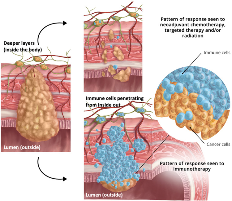
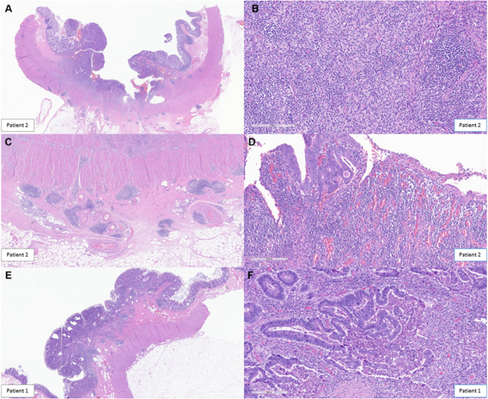
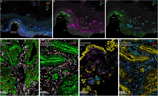
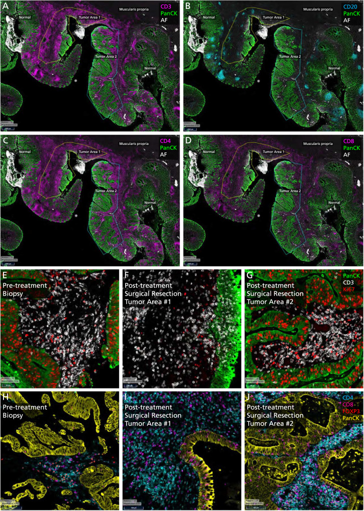
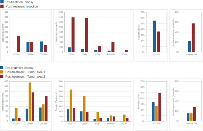
In patients with locally advanced cancer without distant metastases, the neoadjuvant setting presents a platform to evaluate new drugs. For mismatch repair proficient/microsatellite stable (pMMR/MSS) colon and rectal cancer, immunotherapy has shown limited efficacy. Herein, we report exceptional responses observed with neoadjuvant botensilimab (BOT), an Fc-enhanced next-generation anti-CTLA-4 antibody, alongside balstilimab (BAL; an anti-PD-1 antibody) in two patients with pMMR/MSS colon and rectal cancer. The histological pattern of rapid immune response observed ("inside-out" (serosa-to-mucosa) tumor regression) has not been described previously in this setting. Spatial biology analyses (RareCyte Inc.) reveal mechanisms of actions of BOT, a novel innate-adaptive immune activator. These observations have downstream implications for clinical trial designs using neoadjuvant immunotherapy and potentially sparing patients chemotherapy.


Conflict of interest statement

Author PMK reports grants paid to the institution by Merck, Agenus Bio, Novartis, Advanced Accelerator Applications, Tersera, and Boston Scientific; a consultancy and advisory board relationship with Elicio (scientific advisory board member/shares/stock ownership); consultancy/advisory board fees from Guardant Health, Natera, Foundation Medicine, Illumina, BostonGene, Merck/MSD Oncology, Tempus, Bayer, Lilly, Delcath Systems, IPBA, QED Therapeutics, Boston Healthcare Associates, Servier, Taiho Oncology, Exact Sciences, Daiichi Sankyo/AstraZeneca, Eisai, Saga Diagnostics, Neogenomics, Do More Diagnostics AS, and Seattle Genetics; consulting fees paid to the institution by Taiho Pharmaceutical and Ipsen; receiving travel support from AstraZeneca for presentation of an investigator initiated trial. All other authors report no COIs. MH is a member of the board of directors for Bristol Myers Squibb; is a founder of Champions Oncology, Inc., and Nelum Inc.; holds stock options for Champions Oncology, Inc.; InxMed; Biooncotech; and Bristol Myers Squibb; has received research support from Agenus; has received honoraria from InxMed, Oncomatrix, MiNKi, and Peaches Biotech; and has received royalties from Myriad and Khar. MDJ has received honorarium as a speaker for Covidien. A.P. has received honorarium as a consultant for Medtronic, Vioptix, Ethicon, and Intuitive. MAS reports receiving research funding from Astellas Pharma Inc., Merck, Bristol Myers Squibb and Oncolys BioPharma and serving a leadership or judiciary role in board, society, committee or advocacy groups for the American Society of Clinical Oncology Leadership Council. U.K. reports honoraria and travel/accommodations from Cardinal Health, and 2nd.MD. DS reports consultancy for Ciox Health and Zephyr AI, Inc., and speaker/lecturer for Natera, Inc. AJO discloses royalties from Guardant Health and Natera. All other authors have no declared competing interests or financial ties to disclose.

Figures

Fig. 1
Fig. 1. ‘Inside-out’ (serosa-to-mucosa) regression pattern of response seen in patients with colon and rectal cancer receiving botensilimab plus balstilimab in the neoadjuvant setting.
As shown, compared to traditional responses seen with chemotherapy, targeted therapy and/or radiation, the residual proportion of the tumor cells are all confined to the luminal surface with immunotherapy. This can be best described as a wave or tsunami of immune cells infiltration and subsequent cancer cell death.
Fig. 2
Fig. 2. Histopathological Rreview outlining key features noted post-treatment with immunotherapy.
Histopathologic review was based on surgical specimens of the tumors post-treatment that were entirely submitted for the two patients showing marked therapeutic response. Microscopically, a dense mixed inflammatory infiltrate was identified surrounding the tumor mass in Patient 2 (A). The infiltrate was lymphoplasmacytic-rich but also contained frequent macrophages (some foamy), occasional multinucleated giant cells, eosinophils, and neutrophils (B). Tertiary lymphoid structures (TLS) or Crohn like reaction was commonly seen at the periphery (C). Neovascularization was a prominent feature in most tumor beds, and granulation tissue predominated along the luminal surface (D). A similar response was noted in Patient 1. Rather than haphazardly arranged within dense fibrosis, as is often seen with neoadjuvant chemotherapy, viable tumor was often superficially oriented near the luminal surface within the tumor center, with dense inflammation surrounding the periphery and comprising most of the grossly identifiable tumor bed (E). Residual tumor glands often demonstrated evidence of ongoing destruction with incomplete lumens and frequent luminal microabscesses (F).
Fig. 3
Fig. 3. Tissue immune-microenvironment correlates assessed pre- and post-treatment with immunotherapy using RareCyte Inc. for the patient with colon cancer.
A Residual tumor at the tip (colonic mucosa) post-treatment: Invasive margin separates normal colon from the adenocarcinoma and an ulcer. “Inside-out” (serosa-to-mucosa) pattern of regression is evident after treatment with the residual tumor only remaining within the mucosa and superficial submucosa; final path pT1aN0, as opposed to earlier at least T2N1 tumor in this first patient (Patient 1). B, C Extensive immune infiltration post-treatment: The section shows extensive expansion & infiltration of CD3+ T-cells (B) confined to the tumor area in comparison to the adjacent normal colonic tissue after treatment. T-cells surround the tumor cells and extend deep into the muscle layer and the serosal layer. Crohn’s-like reaction is present with many CD20+ B-cells (C) forming follicles specifically in the tumor area & extending into the deeper muscularis propria and serosa (Patient 1). D, E Immune Proliferation (comparison pre- and post-treatment): Ki67 proliferation index for immune cells is markedly increased in the post-treatment surgical resection specimen (58%) compared to the pre-treatment biopsy specimen (23%), especially for T-cells (Patient 1). F, G T-cell density (comparison pre- and post-treatment): T cell density (including all T cell subsets) within the tumor increased dramatically with treatment (1398.4/mm2 for resection vs 190.7/mm2 for biopsy). Treg density also increased (411.0/mm2 for resection vs 56.1/mm2 for biopsy). However, the ratio of Treg to effector T helper cells in the tumor decreased (36% for resection vs 55% for biopsy) (Patient 1).
Fig. 4
Fig. 4. Tissue immune-microenvironment correlates assessed pre- and post-treatment with immunotherapy using RareCyte Inc. for the patient with rectal cancer.
CD3+ (A) and CD20+ cells (B) in tumor microenvironment post-treatment: This section shows extensive expansion & infiltration of CD3+ T-cells (A) in tumor area #1 associated with tumor regression upon treatment, in comparison to tumor area #2 which shows less extensive T cell infiltration and no obvious tumor regression (Patient 2). B shows the CD20+ B-Cells and a Crohn’s like reaction. C, D CD4+ and CD8+ cells in tumor microenvironment post-treatment: This section shows: CD4+ T-helper cells are the main immune cell type in the tumor microenvironment (area #1: 1021.8 /mm2; area #2: 389.3) in the post-treatment resection (C). Few T-helper cells are present superficially in the lamina propria of the adjacent normal colonic tissue. CD8 cytotoxic cells (CTLs) are more prevalent within the tumor area #1 (378.4 /mm2) compared to tumor area #2 (134.2 /mm2) consistent with tumor regression in the former but not latter (D). CTLs are markedly increased and clustered in the invasive margin (862.1 /mm2 and 604.1 /mm2) for areas 1 and 2, respectively protecting the normal deeper tissue from invasion by tumor cells (Patient 2). EG Immune Proliferation (comparison pre- and post-treatment): Ki67 proliferation index for immune cells is markedly increased in post-treatment surgical resection tumor area #2 (35%, right, mucosal adenocarcinoma—(G)) compared to area #1 (20%, center, deep invasive adenocarcinoma associated with tumor regression—(F)) and to the pre-treatment biopsy specimen (16%, E) (Patient 2). Note: The regions of interest (ROIs) Tumor Area#1 and Tumor Area #2 are illustrated in (A, B). H, I and J T Cell Density: T cell density after treatment is increased within the tumor area #1 (1267.7/mm2, deep invasive adenocarcinoma associated with tumor regression, I) but not tumor area #2 (543.6/mm2, mucosal adenocarcinoma, J) compared to the pre-treatment biopsy specimen (483.9/mm2, H). Treg density increased with treatment (236.5/mm2 for area #1, 201.3/mm2 for area #2 vs 132.5/mm2 for biopsy). However, the ratio of Treg to effector T helper cells in the tumor decreased for area #1 and increased for area #2 (27% and 49% for resection vs 34% for biopsy) (Patient 2). Note: The regions of interest (ROIs) Tumor Area #1 and Tumor Area #2 are illustrated in (A, B).
Fig. 5
Fig. 5. Quantitative changes in immune cell populations noted pre- and post-treatment with immunotherapy.
A Graphs showing changes in immune cell populations in Patient 1 with colon cancer (Top Panel - Patient 1): Graphs showing changes in immune cell populations in Patient 1: All B and T cell densities increase in the resection samples (red) compared to the pre-treatment biopsy (blue) while macrophage populations decreased. Proportion of Th (FOXP3+) cells that are Tregs decreased in the resection samples compared to the pre-treatment biopsy. Finally, the proportion of immune cells that are proliferating (Ki67+) is increased in the resection samples compared to the pre-treatment biopsy. B Graphs showing changes in immune cell populations in Patient 2 with rectal cancer (Lower Panel - Patient 2): All B and T cell densities increase in tumor area #1 of resection samples (yellow) compared to tumor area #2 (red) and pre-treatment biopsy (blue); macrophage populations increased in both tumor areas with treatment. Proportion of Th cells that are Treg (FOXP3+) decreased in tumor area #1 and increased in tumor area #2 of the resection samples compared to the pre-treatment biopsy. Finally, the proportion of immune cells that are proliferating is increased in tumor area #2 of the resection samples compared to the pre-treatment biopsy.

References

    1. Le Saux O, Lounici Y, Wajda P, Barrin S, Caux C, Dubois B, et al. Neoadjuvant immune checkpoint inhibitors in cancer, current state of the art. Crit Rev Oncol Hematol. 2021;157:103172. doi: 10.1016/j.critrevonc.2020.103172. - DOI - PubMed
    1. Marron TU, Galsky MD, Taouli B, Fiel MI, Ward S, Kim E, et al. Neoadjuvant clinical trials provide a window of opportunity for cancer drug discovery. Nat Med. 2022;28:626–9. doi: 10.1038/s41591-022-01681-x. - DOI - PMC - PubMed
    1. Chalabi M. The promise of neoadjuvant immunotherapy across solid tumors. 2023. https://meetings.asco.org/2023-asco-annual-meeting/15057?presentation=22... (Accessed 18 Aug 2023).
    1. El-Khoueiry AB, Fakih M, Gordon MS, Tsimberidou AM, Bullock AJ, Wilky BA, et al. Results from a phase 1a/1b study of botensilimab (BOT), a novel innate/adaptive immune activator, plus balstilimab (BAL; anti-PD-1 antibody) in metastatic heavily pretreated microsatellite stable colorectal cancer (MSS CRC) J Clin Oncol. 2023;41:LBA8. doi: 10.1200/JCO.2023.41.4_suppl.LBA8. - DOI
    1. Delepine C, Levey D, Krishnan S, Kim K-S, Sonabend A, Wilkens M, et al. 470 Botensilimab, an Fc-enhanced CTLA-4 antibody, enhances innate and adaptive immune activation to promote superior anti-tumor immunity in cold and I-O refractory tumors. J Immunother Cancer. 2022;10:A490.

MeSH terms

Substances