. 2023 Sep 4:14:1231352.
doi: 10.3389/fphys.2023.1231352. eCollection 2023.

CD51 labels periosteal injury-responsive osteoprogenitors

Affiliations

CD51 labels periosteal injury-responsive osteoprogenitors

Ye Cao et al. Front Physiol. .

Abstract

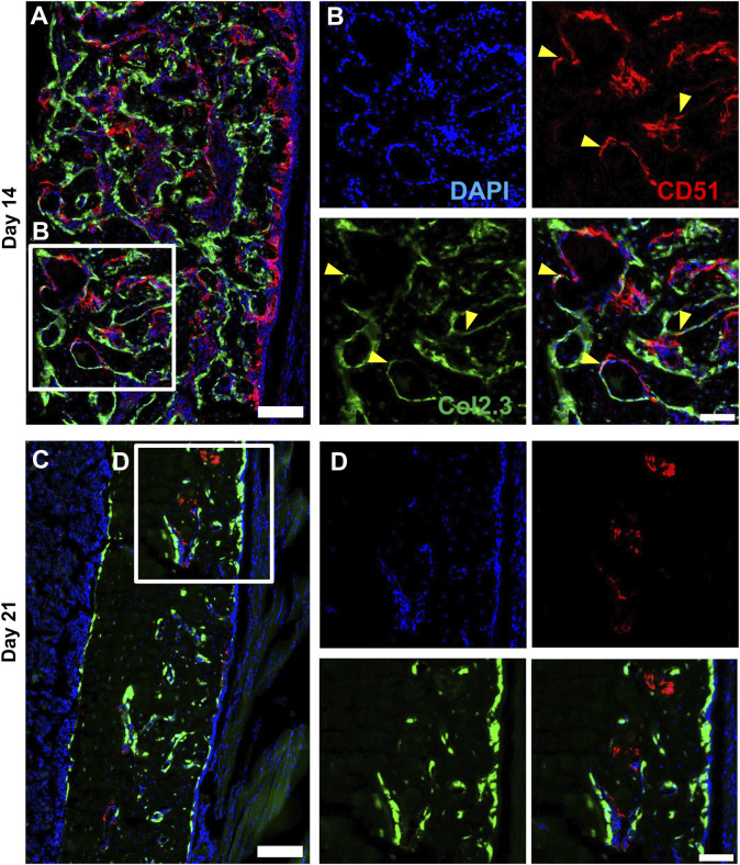
The periosteum is a critical source of skeletal stem and progenitor cells (SSPCs) that form callus tissue in response to injury. There is yet to be a consensus on how to identify SSPCs in the adult periosteum. The aim of this study was to understand how potential murine periosteal SSPC populations behave in vivo and in response to injury. We evaluated the in vivo differentiation potential of Sca1-CD51+ and Sca1+CD51+ cells following transplantation. In vitro, the Sca1+CD51+ population appears to be more primitive multipotent cells, but after transplantation, Sca1-CD51+ cells showed superior engraftment, expansion, and differentiation into chondrocytes and osteoblasts. Despite representing a clear population with flow cytometry, we identified very few Sca1+CD51+ cells histologically. Using a periosteal scratch injury model, we successfully mimicked the endochondral-like healing process seen in unstable fractures, including the expansion and osteochondral differentiation of αSMA+ cells following injury. CD51+ cells were present in the cambium layer of resting periosteum and expanded following injury. Sca1+CD51- cells were mainly localized in the outer periosteal layer. We found that injury increased colony-forming unit fibroblast (CFU-F) formation in the periosteum and led to rapid expansion of CD90+ cells. Several other populations, including Sca1-CD51+ and CD34+ cells, were expanded by day 7. Mice with enhanced fracture healing due to elevated Notch signaling mediated by NICD1 overexpression showed significant expansion of CD51+ and CD34hi cells in the early stages of healing, suggesting these populations contribute to more rapid healing. In conclusion, we demonstrate that periosteal injury leads to the expansion of various SSPC populations, but further studies are required to confirm their lineage hierarchy in the adult skeletal system. Our data indicate that CD51+ skeletal progenitor cells are injury-responsive and show good engraftment and differentiation potential upon transplantation.

Keywords: CD34; CD51; Notch; SCA1; fracture; periosteum.


Conflict of interest statement

The authors declare that the research was conducted in the absence of any commercial or financial relationships that could be construed as a potential conflict of interest.

Figures

FIGURE 1
FIGURE 1
Periosteal Sca1CD51+ cells contribute to osteoblasts and chondrocytes in ectopic bone (A) Experimental design. Periosteal donor cells were isolated from CAG-Tom/Col2.3GFP animals. Sorted populations (4,900–8,000 cells) were mixed with 750,000 bone marrow stromal cells (BMSCs) from wild type animals. After 4 weeks, implants were extracted. Representative image of BMSC only ossicle is shown. Figure partially created with BioRender. (B) X-rays of representative ossicles formed from the Sca1CD51+, and Sca1+CD51+ cells. Representative sections showing cells derived from sorted (C) Sca1CD51+ and (D) Sca1+CD51+ populations in ossicles. Magnified images indicating the red Tom+ donor cells (red arrowheads), and yellow donor cell-derived osteoblasts (yellow arrowheads) and chondrocytes (blue arrowheads) are shown in (i,ii). Sections were counterstained with DAPI. (E) tdTomato+ (donor), and (F) Col2.3GFP+ periosteum and (where present) endosteum surface was calculated (n = 4–5 implants/group). **p < 0.01 (t-test). Tom, tdTomato; DAPI, 4′,6-diamidino-2-phenylindole. Scale bars are 200 µm (C,D), and 50 µm (i, ii).
FIGURE 2
FIGURE 2
Time course of periosteal response to scratch injury. (A) Experimental design for histology and flow analysis of αSMACreER/Tom/Col2.3GFP mice following periosteal injury, created with BioRender. (B) Brightfield imaging of safranin O and fast green stained femur sections showing periosteal response following local injury at different time points (n = 3–5). BM, bone marrow; CB, cortical bone; Peri, periosteum (injured periosteum and healing response); M, muscle. Scale bars are 200 µm.
FIGURE 3
FIGURE 3
Alpha smooth muscle actin (αSMA) identifies injury-responsive periosteal stem and progenitor populations. Representative histology showing periosteum injury response compared to the uninjured femur at day 3 (n = 3), 7 (n = 4), 14 (n = 3), 21 (n = 4), and 28 (n = 2) following injury in αSMACreER/Tom/Col2.3GFP mice. DAPI (white), αSMA (red), Col2.3 (green) were labelled. αSMA cells rapidly expanded as soon as the injury occurred, these cells contributed to periosteum healing by giving rise to Col2.3GFP labelled osteoblasts (yellow arrowheads). BM, bone marrow; CB, cortical bone; Peri, periosteum (injured periosteum and healing response); M, muscle. Scale bars are 200 µm. DAPI, 4′,6-diamidino-2-phenylindole.
FIGURE 4
FIGURE 4
Expansion of cells expressing markers including CD90 and CD34 occurs after injury. αSMACreER/Tom/Col2.3GFP mice were treated with tamoxifen at day −1 and day 0, and had periosteal cells isolated 3 and 7 days later, uninjured αSMACreER/Tom/Col2.3GFP mice were treated with tamoxifen 1 and 2 days before harvesting. (A) The frequency of CD45/Ter119/CD31 (Lin) cells (n = 6–8). Expression of cell surface markers with low (B), and high (C) expression in the periosteum following injury (n = 5-6). (D) Expression of populations expressing Sca1 and CD51 in a separate cohort of B6 mice (n = 3). *p < 0.05 compared to uninjured, #p < 0.05 compared to day 3 with one way ANOVA followed by Tukey’s post hoc test.
FIGURE 5
FIGURE 5
CD51+ periosteal cells expand in response to local injury. Representative histology showing periosteum injury response compared to the uninjured femur at day 3 (n = 3), 7 (n = 4), 14 (n = 3), 21 (n = 4), and 28 (n = 2) following injury. Sca1+ cells mainly resided in the outer layer of the periosteum and did not contribute much to healing; CD51 cells localized in the inner layer of the periosteum, contributed to bone and periosteum formation. Sca1+CD51+ cells (yellow arrowheads) were rare without injury and may decrease with injury. BM, bone marrow; CB, cortical bone; Peri, periosteum (injured periosteum and healing response). Scale bars are 200 µm. DAPI, 4′,6-diamidino-2-phenylindole.
FIGURE 6
FIGURE 6
CD51 expression is detectable in some osteoblasts during active bone formation. Representative histology showing the localization of CD51 in relation to Col2.3GFP+ osteoblasts on day 14 (A,B) and day 21 (C,D) following injury (n = 3–4). DAPI (white), Col2.3 (green), CD51 (red) were labelled. CD51 labelled osteoblasts (yellow arrowheads) were found on day 14 post injury, but these cells disappeared on day 21. Scale bars are 200 µm (A,C) or 100 µm (B,D). DAPI, 4′,6-diamidino-2-phenylindole.
FIGURE 7
FIGURE 7
CD34hi cells are stimulated with NICD1 overexpression following periosteal injury. (A) Experimental design of flow cytometry analysis on αSMACreER/NICD1 animals following periosteal injury (n = 4–5), partially created with BioRender. (B) The proportion of hematopoietic lineage negative (Lin) from WT (Cre) and NICD1+ (Cre+) mice, representative flow plot shown in (C). (D) The proportion of single marker expression within the Lin fraction. (E) The proportion of Sca1/CD51 populations within Lin. (F) Representative flow plots showing gating strategy of CD34med and CD34hi populations, and Sca1/CD51 populations within periosteal Lin of WT and NICD1+ mice. (G) The frequency of Sca1/CD51 populations within CD34hi cells. (H) The frequency of Sca1/CD51 populations within CD34med cells. (I) Representative flow plots showing Sca1/CD51 populations within CD34hi and CD34med populations of WT and NICD1+ mice. *p < 0.05 compared to WT with unpaired t-test. Percentages are specific to the sample. WT, wild type; NICD1, Notch intracellular domain 1.
FIGURE 8
FIGURE 8
Injury enhances CFU-F formation overall and in CD34+ cells. Unilateral periosteal injury was performed on WT mice, and periosteal cells were harvested 3 days after injury, the uninjured cells were isolated from the matched uninjured legs (n = 3). (A) Representative plate image showing CFU-F of periosteal Lin-populations from uninjured and day 3 injury, stained with crystal violet, and (B) quantification of CFU-F for each population. CFU-Fs from the CD34+ cells were differentiated with dual-lineage media, and stained with alkaline phosphate (ALP, for osteogenesis), and oil red O (ORO, for adipogenesis), and the stained colonies were quantified (C), representative stained colonies are shown in (D). (E,F) Unilateral periosteal injury was performed on αSMACreER/NICD1 mice as indicated in Figure 7A, and periosteal cells sorted at day 3. (E) Quantification of CFU-F for CD34 populations in WT and NICD1+ animals (n = 2–3), and representative plate images with crystal violet staining of CFU-Fs are shown in (F). Two-way ANOVA with Tukey’s post hoc test: *p < 0.05 compared to uninjured, #p < 0.05 compared to Lin within the same time point. Lin: hematopoietic lineage negative.

References

    1. Abdallah B. M., Al-Shammary A., Skagen P., Abu Dawud R., Adjaye J., Aldahmash A., et al. (2015). CD34 defines an osteoprogenitor cell population in mouse bone marrow stromal cells. Stem Cell. Res. 15 (3), 449–458. 10.1016/j.scr.2015.09.005 - DOI - PubMed
    1. Ball M. D., Bonzani I. C., Bovis M. J., Williams A., Stevens M. M. (2011). Human periosteum is a source of cells for orthopaedic tissue engineering: a pilot study. Clin. Orthop. Relat. Res. 469 (11), 3085–3093. 10.1007/s11999-011-1895-x - DOI - PMC - PubMed
    1. Baryawno N., Przybylski D., Kowalczyk M. S., Kfoury Y., Severe N., Gustafsson K., et al. (2019). A cellular taxonomy of the bone marrow stroma in homeostasis and leukemia. Cell. 177 (7), 1915–1932. 10.1016/j.cell.2019.04.040 - DOI - PMC - PubMed
    1. Cao Y., Bolam S. M., Boss A. L., Murray H. C., Dalbeth N., Brooks A. E. S., et al. (2022). Characterization of adult human skeletal cells in different tissues reveals a CD90+CD34+ periosteal stem cell population. Cold Spring Harbor Laboratory. - PubMed
    1. Cao Y., Buckels E. J., Matthews B. G. (2020). Markers for identification of postnatal skeletal stem cells in vivo . Curr. Osteoporos. Rep. 18 (6), 655–665. 10.1007/s11914-020-00622-2 - DOI - PubMed

LinkOut - more resources