. 2023 Aug 31;26(10):107803.
doi: 10.1016/j.isci.2023.107803. eCollection 2023 Oct 20.

Oxygenolytic sulfoquinovose degradation by an iron-dependent alkanesulfonate dioxygenase

Affiliations

Oxygenolytic sulfoquinovose degradation by an iron-dependent alkanesulfonate dioxygenase

Zonghua Ye et al. iScience. .

Abstract

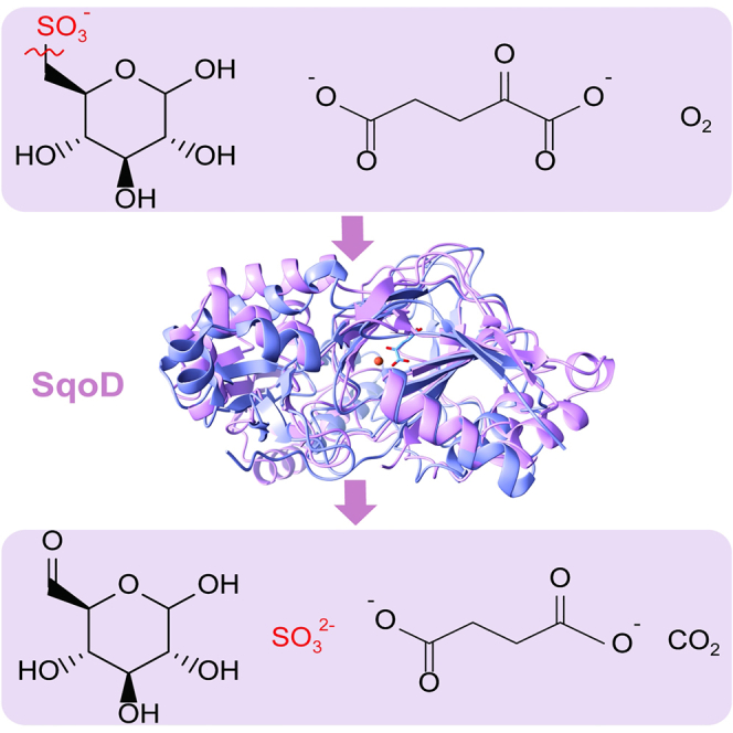
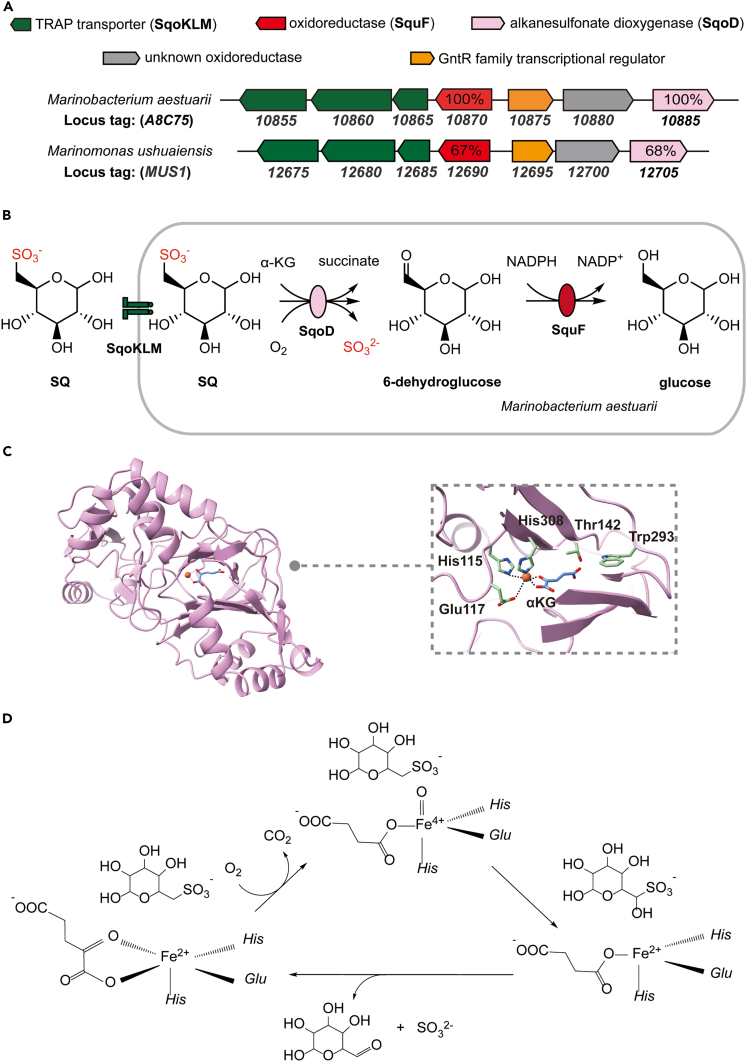
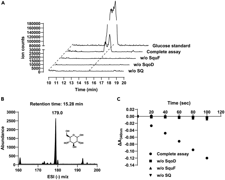
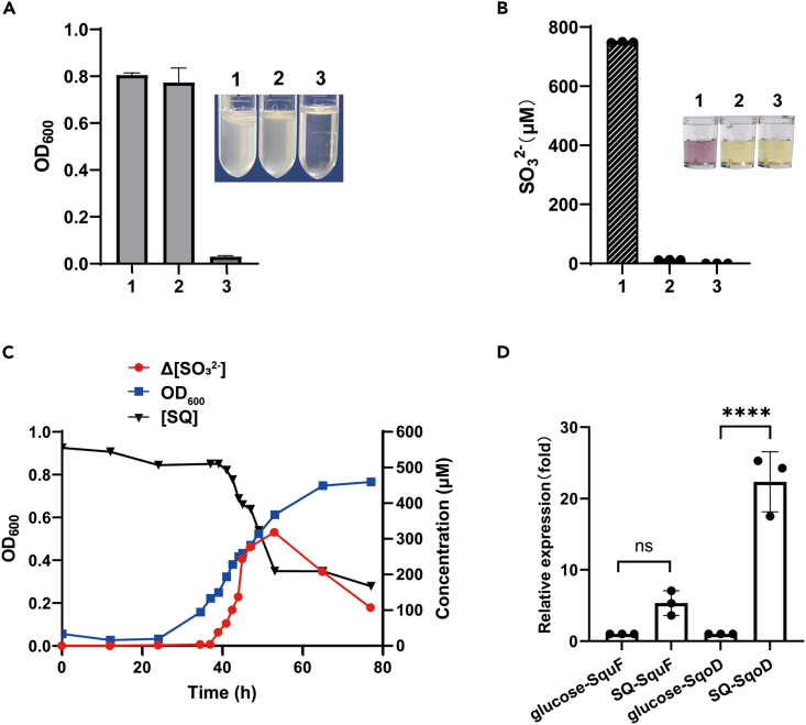
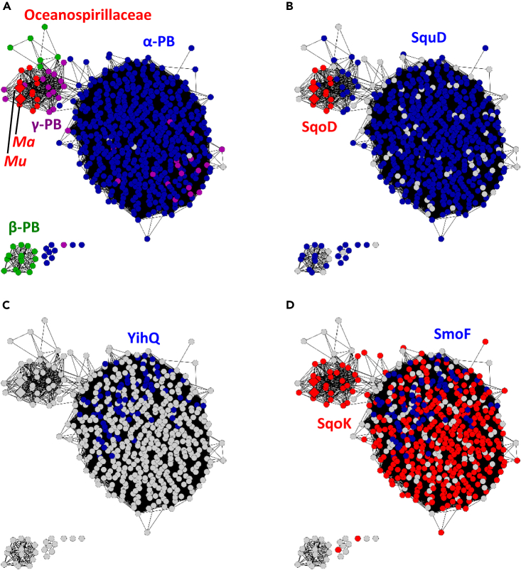
Sulfoquinovose (6-deoxy-6-sulfo-D-glucose, SQ), the polar head group of sulfolipids in plants, is abundant in nature. Many bacteria degrade SQ through pathways termed sulfoglycolysis producing C3 or C2 sulfonates, while certain bacteria degrade SQ through direct oxygenolytic cleavage of the SQ C-S bond, catalyzed by a flavin-dependent alkanesulfonate monooxygenase (sulfo-ASMO pathway). Here we report bioinformatics and biochemical studies revealing an alternative mechanism for oxygenolytic cleavage of the SQ C-S bond, catalyzed by an iron and α-ketoglutarate-dependent alkanesulfonate dioxygenase (SqoD, sulfo-ASDO pathway). In both the ASMO and ASDO pathways, the product 6-dehydroglucose is reduced to glucose by NAD(P)H-dependent SquF. Marinomonas ushuaiensis, a marine bacterium, which harbors the sulfo-ASDO gene cluster is shown utilizing SQ as a carbon source for growth, accompanied by increased transcription of SqoD. The sulfo-ASDO pathway highlights the range of microbial strategies for degradation of this ubiquitous sulfo-sugar, with potential implications for sulfur recycling in different biological environments.

Keywords: Biochemistry; Bioinformatics; Microbial metabolism.


Conflict of interest statement

The authors declare no competing interests.

Figures

None
Graphical abstract
Figure 1
Figure 1
Reaction scheme and gene cluster for the proposed sulfo-ASDO pathway (A) Gene cluster for the sulfo-ASDO pathway in M. aestuarii and M. ushuaiensis. (B) The proposed sulfo-ASDO pathway diagram. (C) AlphaFold model of the active site of SqoD (UniProt A0A1A9EZ58), in which the positions of the Fe and α-ketoglutarate were estimated by overlaying with the crystal structure of E. coli TauD (PDB 1OS7). (D) Reaction scheme for SqoD. See also Figures S2, S6, and S7 and Table S1.
Figure 2
Figure 2
Detection of sulfite formation in the MaSqoD -catalyzed SQ cleavage by a colorimetric Fuchsin assay (A) UV-vis absorption spectra of the complete assay and the negative controls omitting MaSqoD or SQ are shown in red, blue, and black, respectively. (B) Absorbance at 580 nm of each assay. Inset: Photographs of reaction mixtures. The complete assay 1, the negative control omitting SQ 2, and the negative control omitting SqoD 3. Data are represented as mean ± SD. See also Figures S3 and S4.
Figure 3
Figure 3
LC-MS detection of the SqoD reaction product 6-dehydroglucose (A) HPLC elution profiles of the DNPH derivatized products of the SqoD reaction, monitoring the absorbance at 360 nm, showing product peaks for 1 and 2 in the full assay, corresponding to two stereoisomers of DNPH-6-dehydroglucose. (B) Extracted ion chromatograms (m/z (−) 356.9, the predicted mass of the DNPH-6-dehydroglucose monoanion), showing the same two product peaks 1 and 2. (C–E) The ESI m/z (−) spectrum of the region spanned by three peaks in Figure 3B, showing a species with m/z (−) 356.9 corresponding to different isomers of DNPH-6-dehydroglucose (two possible isomers are depicted). See also Figure S5.
Figure 4
Figure 4
In vitro reconstitution of the sulfo-ASDO pathway showing the formation of glucose (A) Extracted ion chromatograms (m/z (−) 179.0, the predicted mass of glucose monoanion), showing product peaks in the full assay coeluting with glucose standard. (B) ESI (−) m/z spectrum of the product peak showing a species with m/z (−) 179.0 corresponding to glucose. (C) Enzyme activity assay monitoring NADPH consumption accompanying 6-dehydroglucose reduction by MaSquF. See also Figure S1.
Figure 5
Figure 5
Growth of M. ushuaiensis in minimal medium containing different carbon sources (A) Tubes 1, 2, and 3 show cells grown in media with SQ, glucose or no additional carbon source. Detect the optical density at 600 nm. Data are represented as mean ± SD. (B) Quantification of sulfite formed in cultures 1, 2, and 3 by a colorimetric fuchsin assay. Inset: Photographs of the respective assays. (C) Optical density of M. ushuaiensis culture (blue) and concentration of SQ ([SQ]) (black) and change in concentration of sulfite (Δ[sulfite]) (red), with respect to time. (D) qPCR analyses of the transcription levels of SqoD and SquF. The transcriptional levels of genes of interest were normalized by that of the 16S rRNA. The induction by SQ was displayed in comparison with the transcriptional data from glucose-grown cells. The error bars represent the standard deviation of three individual experiments. Data are represented as mean ± SD. Significance was assessed by an ordinary one-way ANOVA test (∗∗∗∗p value <0.0001). See also Figures S3 and S8 and Table S2.
Figure 6
Figure 6
SSN of close homologs of glucose-6-dehydrogenase SquF within UniRef50_A0A0K0Y4B2 (A) Sequences present in α-proteobacteria (blue), β-proteobacteria (green), γ-proteobacteria in the family Oceanospirillaceae (red, Mu = Marinomonas ushuaiensis, Ma = Marinobacterium aestuarii), and other γ-proteobacteria (purple). (B) Sequences associated with SquD (blue, sulfo-ASMO pathway) or SqoD (red, sulfo-ASDO pathway), within a 10-ORF window. (C) Sequences associated with sulfoquinovosidase YihQ. (D) Sequences associated with the sulfoquinovoside transporter substrate-binding subunit SmoF (UniRef50_I9XG35), or the putative sulfoquinovose transporter substrate-binding subunit SqoK (UniRef50_V9WJD9).

References

    1. Benning C. Biosynthesis and function of the sulfolipid sulfoquinovosyl diacylglycerol. Annu. Rev. Plant Physiol. Plant Mol. Biol. 1998;49:53–75. doi: 10.1146/annurev.arplant.49.1.53. - DOI - PubMed
    1. Goddard-Borger E.D., Williams S.J. Sulfoquinovose in the biosphere: occurrence, metabolism and functions. Biochem. J. 2017;474:827–849. doi: 10.1042/bcj20160508. - DOI - PubMed
    1. Benson A.A. In: Advances in Lipid Research. Paoletti R., Kritchevsky D., editors. Elsevier; 1963. The Plant Sulfolipid; pp. 387–394. - DOI
    1. Harwood J.L., Nicholls R.G. The plant sulpholipid-- a major component of the sulphur cycle. Biochem. Soc. Trans. 1979;7:440–447. doi: 10.1042/bst0070440. - DOI - PubMed
    1. Sanda S., Leustek T., Theisen M.J., Garavito R.M., Benning C. Recombinant Arabidopsis SQD1 converts UDP-glucose and sulfite to the sulfolipid head group precursor UDP-sulfoquinovose in vitro. J. Biol. Chem. 2001;276:3941–3946. doi: 10.1074/jbc.M008200200. - DOI - PubMed

LinkOut - more resources