

Median nerve versus flexor tendons: visualization of median nerve level changes in the proximal carpal tunnel during wrist movement with dynamic high-resolution ultrasound
- PMID: 37732110
- PMCID: PMC10508267
- DOI: 10.15557/jou.2023.0020
Median nerve versus flexor tendons: visualization of median nerve level changes in the proximal carpal tunnel during wrist movement with dynamic high-resolution ultrasound
Abstract
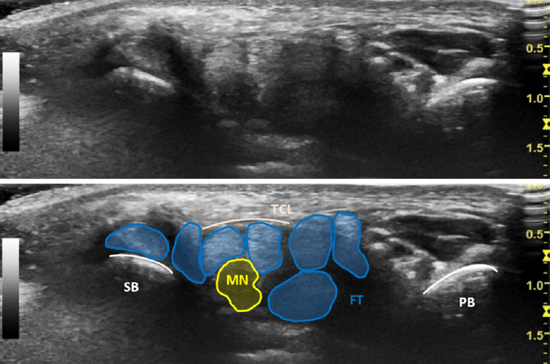
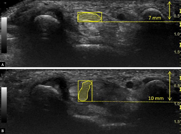
Aim: The purpose of this prospective ultrasound study was to document dorso-palmar (vertical) displacement of the median nerve in relation to the superficial flexor tendons at the level of the carpal tunnel. Furthermore, the gliding patterns of the median nerve were characterized. The presence of vertical gliding was intended to serve as an additional bio-kinematic parameter of median nerve movement, and will be referred to as a 'level change'.
Material and methods: In this study, a total of 32 healthy young individuals underwent dynamic high-resolution ultrasound examinations of both wrists. The neutral position, and maximum flexion and extension of the wrist had to be reached in active and passive movement. The gliding patterns were determined in relation to the superficial flexor tendons. When no vertical nerve gliding was observed, it was characterized as 'no level change'.
Results: The presence of a level change prevailed in the healthy young cohort and was observed in 84% (27/32) of individuals during wrist flexion. The following gliding pattern was distinctively the most common: gliding of the entire nerve in between the flexor tendons in active but not in passive movement of the right and left wrists (13/27; 48%). The extent of vertical displacement was found to be associated with the gliding pattern (Kruskal-Wallis test).
Conclusions: Movement in the carpal tunnel allows the median nerve to adapt to biomechanical stress. Dynamic ultrasound can demonstrate median nerve level changes in response to wrist movements. Furthermore, a typical gliding pattern was characterized. The presence of level change and gliding patterns were proposed as additional movement parameters during wrist flexion in healthy individuals.
Keywords: carpal tunnel; dynamic high-resolution ultrasound; median nerve gliding; nerve biomechanics.
© 2023 Suren Armeni Jengojan et al., published by Sciendo.
Conflict of interest statement
Conflict of interest All authors declare no conflicts of interest. No funding was received for this study.
Figures


Similar articles
-
Transverse Ultrasound Assessment of the Displacement of the Median Nerve in the Carpal Tunnel during Wrist and Finger Motion in Healthy Volunteers.J Nippon Med Sch. 2015;82(4):170-9. doi: 10.1272/jnms.82.170. J Nippon Med Sch. 2015. PMID: 26328793
-
Effects of wrist extension on median nerve and flexor tendon excursions in patients with carpal tunnel syndrome: a case control study.BMC Musculoskelet Disord. 2021 May 24;22(1):477. doi: 10.1186/s12891-021-04349-8. BMC Musculoskelet Disord. 2021. PMID: 34030693 Free PMC article.
-
Anatomic relations between the median nerve and flexor tendons in the carpal tunnel: MR evaluation in normal volunteers.AJR Am J Roentgenol. 1989 Sep;153(3):533-6. doi: 10.2214/ajr.153.3.533. AJR Am J Roentgenol. 1989. PMID: 2763951
-
Median nerve movement in the carpal tunnel before and after carpal tunnel release using transverse ultrasound.J Orthop Surg (Hong Kong). 2017 Sep-Dec;25(3):2309499017730422. doi: 10.1177/2309499017730422. J Orthop Surg (Hong Kong). 2017. PMID: 28920545
-
Interfascicular Gliding Dysfunction Relation with Focal Neuropathy in Diabetic Patients with Carpal Tunnel Syndrome.J Hand Microsurg. 2020 Oct 4;14(1):3-9. doi: 10.1055/s-0040-1718236. eCollection 2022 Jan. J Hand Microsurg. 2020. PMID: 35256822 Free PMC article. Review.
Cited by
-
Ultrasonography of the ulnar nerve loop in relation to the flexor carpi ulnaris tendon.J Ultrason. 2024 Nov 30;24(98):1-5. doi: 10.15557/jou.2024.0027. eCollection 2024 Dec. J Ultrason. 2024. PMID: 39619260 Free PMC article.
References
-
- Hara Y, Tajiri Y, Kawano K, Hoshikawa S Evaluation of restricted motion area of the median nerve in patients with carpal tunnel syndrome: a new measurement method using an ultrasonographic video image J Hand Surg Asian Pac Vol 2021;26(4):635. doi: 10.1142/s2424835521500612. : . . ; ( ): –. . doi: . - DOI - PubMed
LinkOut - more resources
Full Text Sources