[Preprint]. 2023 Sep 5:2023.09.05.556368.
doi: 10.1101/2023.09.05.556368.

Pervasive mislocalization of pathogenic coding variants underlying human disorders

Affiliations

Pervasive mislocalization of pathogenic coding variants underlying human disorders

Jessica Lacoste et al. bioRxiv. .

Update in

  • Pervasive mislocalization of pathogenic coding variants underlying human disorders.
    Lacoste J, Haghighi M, Haider S, Reno C, Lin ZY, Segal D, Qian WW, Xiong X, Teelucksingh T, Miglietta E, Shafqat-Abbasi H, Ryder PV, Senft R, Cimini BA, Murray RR, Nyirakanani C, Hao T, McClain GG, Roth FP, Calderwood MA, Hill DE, Vidal M, Yi SS, Sahni N, Peng J, Gingras AC, Singh S, Carpenter AE, Taipale M. Lacoste J, et al. Cell. 2024 Nov 14;187(23):6725-6741.e13. doi: 10.1016/j.cell.2024.09.003. Epub 2024 Sep 30. Cell. 2024. PMID: 39353438

Abstract

Widespread sequencing has yielded thousands of missense variants predicted or confirmed as disease-causing. This creates a new bottleneck: determining the functional impact of each variant - largely a painstaking, customized process undertaken one or a few genes or variants at a time. Here, we established a high-throughput imaging platform to assay the impact of coding variation on protein localization, evaluating 3,547 missense variants of over 1,000 genes and phenotypes. We discovered that mislocalization is a common consequence of coding variation, affecting about one-sixth of all pathogenic missense variants, all cellular compartments, and recessive and dominant disorders alike. Mislocalization is primarily driven by effects on protein stability and membrane insertion rather than disruptions of trafficking signals or specific interactions. Furthermore, mislocalization patterns help explain pleiotropy and disease severity and provide insights on variants of unknown significance. Our publicly available resource will likely accelerate the understanding of coding variation in human diseases.


Conflict of interest statement

DECLARATION OF INTERESTS A.E.C. serves as a scientific advisor for Recursion, which uses image-based profiling and Cell Painting for drug discovery and receives honoraria for occasional talks at pharmaceutical and biotechnology companies. The other authors declare no competing interests relevant to this manuscript.

Figures
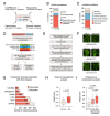
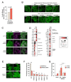
Figure 1.
Figure 1.. Systematic profiling of subcellular localization of missense variants.
A) Sources of variants used in this study. B) Available ClinVar annotations for the variants used in this study. C) Reported inheritance pattern of variants used in this study. D) Pipeline for high-content screen for protein localization. E) Computational pipeline for analyzing localization patterns and comparison of reference alleles and variants. F) Examples of variants with high, medium and low similarity to the reference allele. G) Reference protein localization in this study compared to other large-scale studies. H) Correlation coefficient of localization patterns between reference alleles and missense variants for visually identified hits and non-hits. Statistical significance was calculated with a Mann-Whitney test. I) Correlation coefficient of localization patterns between reference alleles and missense variants for non-hits, low penetrance hits and high penetrance hits. Statistical significance was calculated with ANOVA with Tukey’s correction for multiple testing.
Figure 2.
Figure 2.. Mislocalization map of missense variants.
The overall pattern of mislocalization is shown in the center. Each line represents a mislocalized variant. The color of the line indicates destination compartment (i.e. mislocalization compartment). Examples of mislocalized variants for different categories are shown with manual annotation and the associated disease phenotype. Scale bar, 20 μm.
Figure 3.
Figure 3.. Mislocalization and cellular compartments.
A) Mislocalization affects some genes more than others. Fraction of mislocalized variants for all genes compared to those genes for which already one variant is mislocalized. Statistical significance was calculated with Fisher’s exact test. B) Mislocalization affects some compartments more than others. Relative enrichment of mislocalized variants by localization of the reference protein. Red and blue circles represent compartments from where significantly more (red) or fewer (blue) variants are mislocalized. C) Mislocalized variants are enriched in proteins normally localized to the secretory compartment. Statistical significance was calculated with Fisher’s exact test. D) Examples of mislocalized variants of cytoskeletal proteins. Top, keratin proteins forming distinct punctae. Bottom, mislocalized variants of tubulin and doublecortin, a microtubule-associated protein. E) Examples of missense variants forming distinct foci. Missense variants of SPOP associated with prostate cancer form more foci than the reference protein, whereas coding variants associated with endometrial cancer do not form foci. F) Inheritance pattern of mislocalized variants forming distinct foci and those with other localization patterns. Statistical significance was calculated with Fisher’s exact test. G) Comparison of mislocalization results from this study and from Banani et al. for variants predicted to dysregulate biomolecular condensates. Statistical significance was calculated with Fisher’s exact test with Bonferroni correction for multiple hypotheses.
Figure 4.
Figure 4.. Features associated with mislocalization.
A) Mislocalized variants are enriched in pathogenic and likely pathogenic variants. B) Mislocalized variants are predicted to be more damaging by Polyphen-2. C) Pathogenic and likely pathogenic variants are mislocalized more often than benign or likely benign variants. D) Mutations causing mislocalization are not enriched in post-translational modification sites. E) Mutations causing mislocalization do not disrupt protein-protein interactions more often than mutations leading to normal localization, as assessed by yeast two-hybrid assay and validated by orthogonal assays. F) Mutations causing mislocalization are not enriched in signal peptides. G) Mutations causing mislocalization are highly enriched in transmembrane domains. H) Mislocalized variants interact more with chaperones and quality-control factors than normally localized variants, as determined by LUMIER assays. I-K) Comparison of chaperone and quality control factor interactions of reference proteins and mislocalized or normally localized proteins for Hsp70/HSPA8 (I), Grp78/HSPA5 (J), and Chip/STUB1 (K). Statistical significance was calculated with a chi-square test (panels A and B), Fisher’s exact test (panels D-H), and Mann-Whitney test (panels I-K)
Figure 5.
Figure 5.. Mislocalization and disease phenotypes.
A) Variants of the same gene that have a distinct localization pattern are more often associated with distinct disease phenotypes than variants that are similarly localized. Statistical significance was calculated with Fisher’s exact test. B) Top, loss of membrane localization of PLP1 variants is concordant with disease manifestation. Bottom, loss of intermediate filament staining and appearance of distinct punctae (arrowheads) with GFAP variants correlates with age of onset. C) Distinct localization of beta-actin variants underlying different diseases. Filamentous actin staining with phalloidin (magenta) shows distinct patterns with wild-type actin and each mutant. D) Proximity interactomes of wild-type actin and R183W and E364K variants were determined by BioID in HEK293 cells. The graph shows selected interactions; the full dataset is available in Table S1 E) Mislocalization of SMAD2 D304G variant from the nucleus to the cytoplasm. Wild-type and mutant SMAD2-3xFLAG-V5 constructs were transfected into HeLa and U2OS cells and stained with anti-FLAG antibody (green). F) SMAD2 D304G interacts less with the transcriptional co-regulator SKI and more with the TGFβ receptor TGFBR1. Indicated 3xFLAG-V5 tagged constructs were co-transfected into HEK293T cells with Nanoluc-tagged wild-type SMAD2 or the D304G variant, and interaction was assayed with LUMIER assay. Statistical significance was calculated with ANOVA with Tukey’s correction for multiple hypotheses. ***, p < 0.001, *, p < 0.05. G) SMAD2 D304G is a weaker transactivator than wild-type SMAD2. Indicated constructs were co-transfected with 3TP-lux reporter and Nanoluc control into MDA-231 cells and the cells were treated with vehicle control or TGFβ. Transactivation activity measured with luciferase assay. The ratio between Firefly and Nanoluc luminescence was normalized to EGFP control with vehicle treatment. Statistical significance was calculated with ANOVA with Tukey’s correction for multiple hypotheses. ***, p < 0.001, *, p < 0.05.

References

    1. Tabet D., Parikh V., Mali P., Roth F.P., and Claussnitzer M. (2022). Scalable Functional Assays for the Interpretation of Human Genetic Variation. Annu. Rev. Genet. 56, 441–465. 10.1146/annurev-genet-072920-032107. - DOI - PubMed
    1. Taipale M. (2018). Disruption of protein function by pathogenic mutations: common and uncommon mechanisms. Biochem. Cell Biol. 37, 508. - PubMed
    1. Capriotti E., Ozturk K., and Carter H. (2019). Integrating molecular networks with genetic variant interpretation for precision medicine. WIREs Syst. Biol. Med. 11. 10.1002/wsbm.1443. - DOI - PMC - PubMed
    1. Vihinen M. (2021). Functional effects of protein variants. Biochimie 180, 104–120. 10.1016/j.biochi.2020.10.009. - DOI - PubMed
    1. Taverna D.M., and Goldstein R.A. (2001). Why are proteins marginally stable? Proteins 46, 105–109. - PubMed

Publication types