Review
. 2023 Sep 5:17:1196606.
doi: 10.3389/fnins.2023.1196606. eCollection 2023.

Development of circadian neurovascular function and its implications

Affiliations
Review

Development of circadian neurovascular function and its implications

Jennifer W Mitchell et al. Front Neurosci. .

Abstract

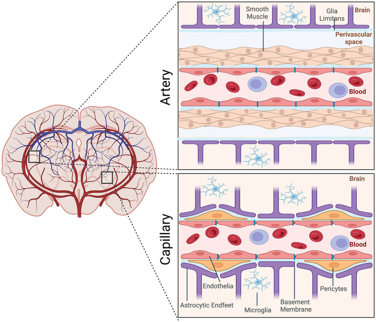
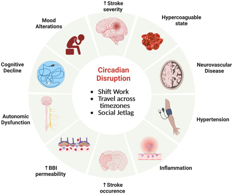
The neurovascular system forms the interface between the tissue of the central nervous system (CNS) and circulating blood. It plays a critical role in regulating movement of ions, small molecules, and cellular regulators into and out of brain tissue and in sustaining brain health. The neurovascular unit (NVU), the cells that form the structural and functional link between cells of the brain and the vasculature, maintains the blood-brain interface (BBI), controls cerebral blood flow, and surveils for injury. The neurovascular system is dynamic; it undergoes tight regulation of biochemical and cellular interactions to balance and support brain function. Development of an intrinsic circadian clock enables the NVU to anticipate rhythmic changes in brain activity and body physiology that occur over the day-night cycle. The development of circadian neurovascular function involves multiple cell types. We address the functional aspects of the circadian clock in the components of the NVU and their effects in regulating neurovascular physiology, including BBI permeability, cerebral blood flow, and inflammation. Disrupting the circadian clock impairs a number of physiological processes associated with the NVU, many of which are correlated with an increased risk of dysfunction and disease. Consequently, understanding the cell biology and physiology of the NVU is critical to diminishing consequences of impaired neurovascular function, including cerebral bleeding and neurodegeneration.

Keywords: blood–brain interface; circadian rhythm disruption; clock; neuroendothelial; tight junctions.


Conflict of interest statement

The authors declare that the research was conducted in the absence of any commercial or financial relationships that could be construed as a potential conflict of interest.

Figures

Figure 1
Figure 1
Schematic representation of the cellular elements of the neurovascular unit (NVU). Elements of the NVU include neuroendothelial cells, mural cells (vascular smooth muscle cells, pericytes), microglia, astrocytic endfeet. The cellular composition differs along the vascular tree. At the level of the artery/arteriole (top), the NVU is composed of neuroendothelial cells making up the inner layer of the vessel wall covered by a thin extracellular basement membrane, ringed by vascular smooth muscle cells, and ensheathed by a glial limitans. The perivascular space containing the cerebrospinal fluid is between pia and the glia limitans formed by astrocytic endfeet. At the capillary level (bottom), the NVU is composed of neuroendothelial cells that share a common basement membrane with pericytes. Pericytes stretch their processes along and around capillaries. Pericytes and endothelial cells are covered by astrocyte endfeet. Created by Biorender.com.
Figure 2
Figure 2
A schematic representation of neurovascular units with cellular elements that regulate cerebral blood flow along the vascular tree. The various types of cells that form the neurovascular unit (NVU) (neurons, astrocytes, mural cells – vascular smooth muscle cells (VSMCs) and pericytes, and neuroendothelium) regulate cerebral blood flow throughout the vascular tree. The cellular composition of the NVU differs along the vascular tree, but the principal cellular components all remain represented, as illustrated here. (A) At the level of penetrating arteries, the NVU is composed of neuroendothelial cells, making up the inner layer of the vessel wall, covered by a thin extracellular basement membrane, ringed by one to three layers of VSMCs, and ensheathed by pia. The Virchow-Robin space containing the cerebrospinal fluid is between the pia and glia limitans formed by astrocytic endfeet. Both VSMCs and astrocytes are innervated by local neurons. (B) Arterioles differ in that (i) there is only one layer of VSMCs, (ii) astrocyte coverage and innervation of the vessel wall and endothelial inner layer are continuous with penetrating arteries and brain capillaries, above and below the arteriole level, respectively. Precapillary arterioles may also contain transitional pericytes, a cell type between pericyte and VSMCs. (C) At the capillary level, the NVU is composed of endothelial cells that share a common basement membrane with pericytes. Pericytes stretch their processes along and around capillaries and make direct, interdigitated or “peg-socket”-like contacts with neuroendothelial cells. Pericytes and neuroendothelial cells are covered by astrocyte endfeet. Both astrocytes and pericytes are innervated by local neurons similar to astrocytes and VSMCs in the upper segments of the vascular tree. Created by Biorender.com.
Figure 3
Figure 3
Molecular circadian clockwork is composed of two interlocking transcription/translation feedback loops (TTFLs). The clock proteins, CLOCK and BMAL1, are integral components of the core circadian timekeeping loop. They form a heterodimer, then induce E-box-mediated transcription of negative regulators, Period (Per1,2,3) and Cryptochrome (Cry1,2) genes. Accumulated PER and CRY proteins repress E-box-mediated transcription until their levels decrease, allowing the cycle to repeat. CLOCK and BMAL1 also control the transcription of the nuclear receptors RORα and REV-ERBα, which modulate BMAL1 mRNA levels by competitive actions on the RRE element residing in the Bmal1 promoter. The cycling of clock components collectively determines the temporal patterning and levels of clock-controlled genes (CCGs), thus generating diverse circadian rhythmic outputs. In addition, a number of signaling molecules, including kinases and ubiquitinases, fine-tune these molecular clock loops. Casein kinase Iε (CKIε) and CKIδ form a complex with PERs and CRYs, phosphorylate (P) PERs and then promote proteasome-dependent degradation of these negative regulators. Phosphorylation of CLOCK facilitates its dimerization with BMAL1 and nuclear entry.
Figure 4
Figure 4
Circadian rhythms are expressed in the various cell types of the NVU. Multiple types of cells comprise the NVU and contribute to neurovascular function. All express near-24-h oscillations in functionality.
Figure 5
Figure 5
Circadian rhythm disfunction adversely effects physiology. Misalignment of circadian clocks between cells, brain regions, tissue, across the organism and the natural world can increase the risk of significant health consequences involving the neurovasculature and brain. Created by Biorender.com.

References

    1. Abbott N. J., Patabendige A. A., Dolman D. E., Yusof S. R., Begley D. J. (2010). Structure and function of the blood-brain barrier. Neurobiol. Dis. 37, 13–25. doi: 10.1016/j.nbd.2009.07.030, PMID: - DOI - PubMed
    1. Abbott N. J., Ronnback L., Hansson E. (2006). Astrocyte-endothelial interactions at the blood-brain barrier. Nat. Rev. Neurosci. 7, 41–53. doi: 10.1038/nrn1824, PMID: - DOI - PubMed
    1. Andreotti F., Davies G. J., Hackett D. R., Khan M. I., De Bart A. C., Aber V. R., et al. . (1988). Major circadian fluctuations in fibrinolytic factors and possible relevance to time of onset of myocardial infarction, sudden cardiac death and stroke. Am. J. Cardiol. 62, 635–637. doi: 10.1016/0002-9149(88)90669-8, PMID: - DOI - PubMed
    1. Andreotti F., Kluft C. (1991). Circadian variation of fibrinolytic activity in blood. Chronobiol. Int. 8, 336–351. - PubMed
    1. Anea C. B., Zhang M., Stepp D. W., Simkins G. B., Reed G., Fulton D. J., et al. . (2009). Vascular disease in mice with a dysfunctional circadian clock. Circulation 119, 1510–1517. doi: 10.1161/CIRCULATIONAHA.108.827477, PMID: - DOI - PMC - PubMed

LinkOut - more resources