. 2023 Oct 31;148(18):1395-1409.
doi: 10.1161/CIRCULATIONAHA.122.061736. Epub 2023 Sep 21.

Combined Treatment of Human Induced Pluripotent Stem Cell-Derived Cardiomyocytes and Endothelial Cells Regenerate the Infarcted Heart in Mice and Non-Human Primates

Affiliations

Combined Treatment of Human Induced Pluripotent Stem Cell-Derived Cardiomyocytes and Endothelial Cells Regenerate the Infarcted Heart in Mice and Non-Human Primates

Yu-Che Cheng et al. Circulation. .

Abstract

Background: Remuscularization of the mammalian heart can be achieved after cell transplantation of human induced pluripotent stem cell (hiPSC)-derived cardiomyocytes (CMs). However, several hurdles remain before implementation into clinical practice. Poor survival of the implanted cells is related to insufficient vascularization, and the potential for fatal arrhythmogenesis is associated with the fetal cell-like nature of immature CMs.

Methods: We generated 3 lines of hiPSC-derived endothelial cells (ECs) and hiPSC-CMs from 3 independent donors and tested hiPSC-CM sarcomeric length, gap junction protein, and calcium-handling ability in coculture with ECs. Next, we examined the therapeutic effect of the cotransplantation of hiPSC-ECs and hiPSC-CMs in nonobese diabetic-severe combined immunodeficiency (NOD-SCID) mice undergoing myocardial infarction (n≥4). Cardiac function was assessed by echocardiography, whereas arrhythmic events were recorded using 3-lead ECGs. We further used healthy non-human primates (n=4) with cell injection to study the cell engraftment, maturation, and integration of transplanted hiPSC-CMs, alone or along with hiPSC-ECs, by histological analysis. Last, we tested the cell therapy in ischemic reperfusion injury in non-human primates (n=4, 3, and 4 for EC+CM, CM, and control, respectively). Cardiac function was evaluated by echocardiography and cardiac MRI, whereas arrhythmic events were monitored by telemetric ECG recorders. Cell engraftment, angiogenesis, and host-graft integration of human grafts were also investigated.

Results: We demonstrated that human iPSC-ECs promote the maturity and function of hiPSC-CMs in vitro and in vivo. When cocultured with ECs, CMs showed more mature phenotypes in cellular structure and function. In the mouse model, cotransplantation augmented the EC-accompanied vascularization in the grafts, promoted the maturity of CMs at the infarct area, and improved cardiac function after myocardial infarction. Furthermore, in non-human primates, transplantation of ECs and CMs significantly enhanced graft size and vasculature and improved cardiac function after ischemic reperfusion.

Conclusions: These results demonstrate the synergistic effect of combining iPSC-derived ECs and CMs for therapy in the postmyocardial infarction heart, enabling a promising strategy toward clinical translation.

Keywords: arrhythmias, cardiac; guided tissue regeneration; induced pluripotent stem cell; models, animal.


Conflict of interest statement

Disclosures None.

Figures

Figure 1.
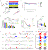
Figure 1.. Co-culture with ECs promotes the maturity of CMs.
A, Sarcomeric structure in EC+CM co-culture and CM alone groups. Scale bars, 20 μm. B, Quantification of sarcomeric length. C, Representative regular calcium transients of CMs co-cultured with ECs or alone. D, Amplitude of calcium transients. E, Slope of calcium upstroke. F, Slope of calcium downstroke. G, Tau decay of calcium transients. H, Maximum diastolic potential of EC+CM co-culture and CM alone groups. I, Ultrastructural images of CM sarcomeric structure (upper panel). Arrowhead, Z disk. Arrow, I band. Ultrastructural images of mitochondria in CMs (lower panel). Scale bars, 1 μm. Statistical significance was determined by two-tailed unpaired t-test in B, D, F and H and determined by the Mann-Whitney test in E and G. n=28 independent samples for EC+CM and n=28 independent samples for CM. *P < 0.05, **P < 0.01, ***P < 0.001, ****P < 0.0001.
Figure 2.
Figure 2.. Co-transplantation of ECs and CMs restores cardiac function and exhibits lower arrhythmic events.
A, Schematic of study design and timeline for cardiac function evaluation. Echocardiography was performed 7 days before surgery, 2 days after surgery and 21 days follow-up. Three-lead electrocardiogram was carried out on day 21, before euthanization. EC+CM, double CM (CM x 2), CM, EC, and vehicle, n=19, 17, 19, 15, and 14 animals, respectively. MI, myocardial infarction. B, Kaplan–Meier analysis over 21 days following myocardial infarction and cell treatment. (Mantel–Cox log-rank test; n=19, 17, 19, 15, and 14 animals for EC + CM, double CM, CM, EC, and vehicle, respectively.) C, Fractional shortening was measured by echocardiography at day −7, day 2 and day 21. D, Overlay of fractional shortening. E, Changes of fractional shortening between day 2 and day 21. n=16, 12, 12, 11, and 11 animals for EC + CM, double CM, CM, EC, and vehicle, respectively in C, D, and E. F, Representative ECG recordings showing premature ventricular contraction (red arrows) and ventricular couplet (blue arrows) in mice. G, Pie chart showing proportion of animals with arrhythmic events. (n=6, 6, 8, 6, and 7 animals for EC+CM, double CM, CM, EC, and vehicle, respectively.) Statistical significance was determined by two-way mixed-effects ANOVA with the Geisser-Greenhouse correction and Tukey’s multiple comparisons test in C and D; and was determined by one-way ANOVA with Tukey’s multiple comparisons test in E. *P < 0.05, **P < 0.01, ***P < 0.001, ****P < 0.0001, ns for not significant.
Figure 3.
Figure 3.. Co-transplantation of ECs exerts trophic effects on the grafts in MI mouse model.
A, Representative images of infarct area stained by picrosirius red and fast green. Scale bar, 1 mm. B, Quantification of infarct area. n=16, 12, 12, 11, and 11 animals for EC+CM, double CM, CM, EC, and vehicle, respectively. C, Representative images of vasculature in the infarct area. Scale bars, 40 μm. D, Quantification of vascular density in the infarct area. n=16, 12, 12, 11, and 11 animals for EC+CM, double CM, CM, EC, and vehicle, respectively. E, Representative whole slide scan images of grafts. Scale bars, 1 mm. F, Engrafted animal ratio in each group. (n=16, 12, and 12 animals for EC+CM, double CM, and CM, respectively.) G, Quantification of graft size. n=15, 9, and 9 animals for EC + CM, double CM, and CM, respectively. H, Representative images of graft vasculature. Scale bars, 40 μm. I, Quantification of vascular density per animal. n=15, 9, and 9 animals for EC + CM, double CM, and CM, respectively. J, Representative images of sarcomere structure of grafts. Scale bars, 5 μm. K, Quantification of sarcomeric length per animal. n=15, 9, and 9 animals for EC + CM, double CM, and CM, respectively. L, Representative images of proliferated cells in grafts stained by Ki67 antibody. Scale bars, 40 μm. M, Quantification of proliferation index per animal. n=15, 9, and 9 animals for EC + CM, double CM, and CM, respectively. Statistical significance was determined by Kruskal-Wallis test with Dunn’s multiple comparison test in B, D and G and determined by one-way ANOVA with Tukey’s multiple comparisons test in I, K, and M. *P < 0.05, **P < 0.01, ***P < 0.001, ns for not significant.
Figure 4.
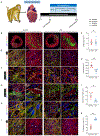
Figure 4.. Co-transplantation of ECs promotes CM maturity in healthy non-human primates.
A, Schematic of study design. B, Representative whole heart slide images of grafts. C, Quantification of graft size. D, Representative images of graft vasculature. Scale bars, 40 μm. E, Quantification of vascular density per animal. F, Representative images of adherens junction expression within grafts. Scale bars, 40 μm. G, Representative images of CM integration with host myocardium. Yellow arrows, pan-cadherin at intercalated disk of CMs. Scale bars, 20 μm. H, Representative images of sarcomeric length in grafts. Scale bars, 10 μm. I, Quantification of sarcomeric length per animal. J, Representative images of proliferated cells in grafts. Scale bars, 40 μm. K, Quantification of proliferation rate per animal. Statistical significance was determined by two-tailed paired t-test in C, E, I, and K. n=4 samples for EC+CM engraftment and n=4 samples for CM engraftment. *P < 0.05, **P < 0.01, ns for not significant.
Figure 5.
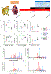
Figure 5.. Co-transplantation of ECs and CMs restores cardiac function after ischemic-reperfusion injury in non-human primates.
A, Schematic of study design. Cardiac magnetic resonance imaging was performed before the day of cell injection and at 28 days follow-up. Echocardiography was performed before I/R surgery, before cell injection and before termination. Electrocardiogram was obtained using a loop recorder throughout the experiment. I/R, ischemic-reperfusion. B, Difference in EF between day 0 and day 28 for each macaque. C, Difference in ESV between day 0 and day 28 for each macaque. D, Difference in EDV between day 0 and day 28 for each macaque. E, Difference in FS between day 0 and day 28 for each macaque. F, Difference in LVESD between day 0 and day 28 for each macaque. G, Difference in LVEDD between day 0 and day 28 for each macaque. n=4 macaques for EC+CM engraftment, n=3 macaques for CM engraftment and n=4 macaques for control. H, Sustained ventricular tachycardia (VT) episodes monitored by loop recorder. I, Sustained VT duration recorded by loop recorder as min per day (m/d). J, Non-sustained VT episodes monitored by loop recorder. K, Non-sustained VT duration recorded by loop recorder as m/d. Statistical significance was determined by one-way ANOVA with Tukey’s multiple comparisons test in B through G.*P < 0.05, **P < 0.01, ns for not significant.
Figure 6.
Figure 6.. Co-transplantation of ECs reduces infarct size in non-human primates with ischemic-reperfusion injury.
A, Representative images of infarct area stained by picrosirius red and fast green. Scale bar, 10 mm. B, Quantification of infarct area. n=4 macaques for EC+CM engraftment, n=3 macaques for CM engraftment, and n=4 macaques for control. C, Representative whole heart slide images of grafts. D, Quantification of graft size. n=4 macaques for EC+CM engraftment and n=3 macaques for CM engraftment. Statistical significance was determined by one-way ANOVA with Tukey’s multiple comparisons test in B and was determined by unpaired t test in D. *P < 0.05, ns for not significant.

References

    1. Laflamme MA, Chen KY, Naumova AV, Muskheli V, Fugate JA, Dupras SK, Reinecke H, Xu C, Hassanipour M, Police S, et al. Cardiomyocytes derived from human embryonic stem cells in pro-survival factors enhance function of infarcted rat hearts. Nat Biotechnol. 2007;25:1015–1024. doi: 10.1038/nbt1327 - DOI - PubMed
    1. Shiba Y, Fernandes S, Zhu WZ, Filice D, Muskheli V, Kim J, Palpant NJ, Gantz J, Moyes KW, Reinecke H, et al. Human ES-cell-derived cardiomyocytes electrically couple and suppress arrhythmias in injured hearts. Nature. 2012;489:322–325. doi: 10.1038/nature11317 - DOI - PMC - PubMed
    1. Romagnuolo R, Masoudpour H, Porta-Sánchez A, Qiang B, Barry J, Laskary A, Qi X, Massé S, Magtibay K, Kawajiri H, et al. Human Embryonic Stem Cell-Derived Cardiomyocytes Regenerate the Infarcted Pig Heart but Induce Ventricular Tachyarrhythmias. Stem Cell Reports. 2019;12:967–981. doi: 10.1016/j.stemcr.2019.04.005 - DOI - PMC - PubMed
    1. Liu Y-W, Chen B, Yang X, Fugate JA, Kalucki FA, Futakuchi-Tsuchida A, Couture L, Vogel KW, Astley CA, Baldessari A, et al. Human embryonic stem cell–derived cardiomyocytes restore function in infarcted hearts of non-human primates. Nature Biotechnology. 2018;36:597–605. doi: 10.1038/nbt.4162 - DOI - PMC - PubMed
    1. Chong JJ, Yang X, Don CW, Minami E, Liu YW, Weyers JJ, Mahoney WM, Van Biber B, Cook SM, Palpant NJ, et al. Human embryonic-stem-cell-derived cardiomyocytes regenerate non-human primate hearts. Nature. 2014;510:273–277. doi: 10.1038/nature13233 - DOI - PMC - PubMed

Publication types