

Mesenchymal Stem Cells Isolated from Equine Hair Follicles Using a Method of Air-Liquid Interface
- PMID: 37733199
- PMCID: PMC10661790
- DOI: 10.1007/s12015-023-10619-w
Mesenchymal Stem Cells Isolated from Equine Hair Follicles Using a Method of Air-Liquid Interface
Abstract
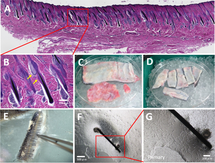
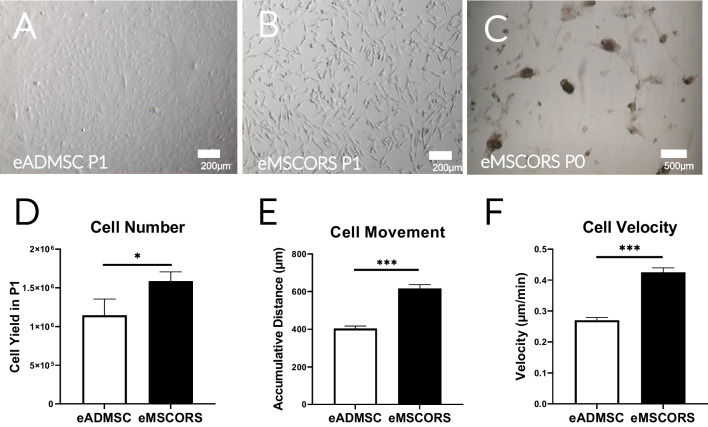
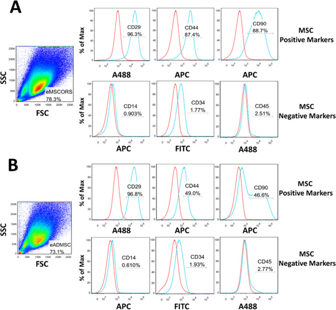
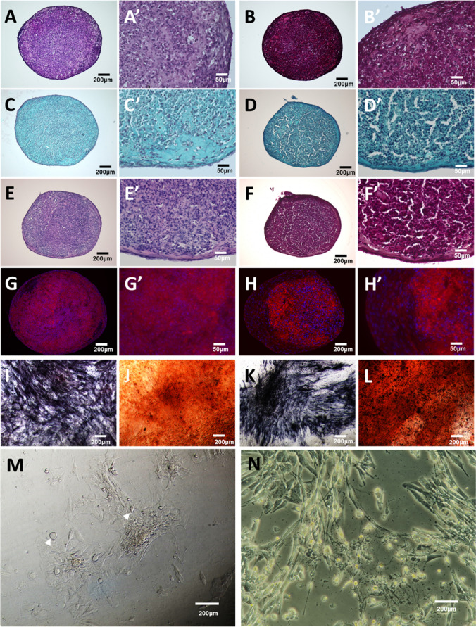
Equine mesenchymal stem cells (MSC) of various origins have been identified in horses, including MSCs from the bone marrow and adipose tissue. However, these stem cell sources are highly invasive in sampling, which thereby limits their clinical application in equine veterinary medicine. This study presents a novel method using an air-liquid interface to isolate stem cells from the hair follicle outer root sheath of the equine forehead skin. These stem cells cultured herewith showed high proliferation and asumed MSC phenotype by expressing MSC positive biomarkers (CD29, CD44 CD90) while not expressing negative markers (CD14, CD34 and CD45). They were capable of differentiating towards chondrogenic, osteogenic and adipogenic lineages, which was comparable with MSCs from adipose tissue. Due to their proliferative phenotype in vitro, MSC-like profile and differentiation capacities, we named them equine mesenchymal stem cells from the hair follicle outer root sheath (eMSCORS). eMSCORS present a promising alternative stem cell source for the equine veterinary medicine.
Keywords: Autologous veterinary therapy; Equine hair follicles; Mesenchymal stem cells; Minimal-invasive cell source; Tri-lineage differentiations.
© 2023. The Author(s).
Conflict of interest statement
The authors declare no conflict of interest.
Figures
Similar articles
-
Characterization and osteogenic potential of equine muscle tissue- and periosteal tissue-derived mesenchymal stem cells in comparison with bone marrow- and adipose tissue-derived mesenchymal stem cells.Am J Vet Res. 2013 May;74(5):790-800. doi: 10.2460/ajvr.74.5.790. Am J Vet Res. 2013. PMID: 23627394
-
Osteogenic Potential of Mesenchymal Stem Cells from Adipose Tissue, Bone Marrow and Hair Follicle Outer Root Sheath in a 3D Crosslinked Gelatin-Based Hydrogel.Int J Mol Sci. 2021 May 20;22(10):5404. doi: 10.3390/ijms22105404. Int J Mol Sci. 2021. PMID: 34065598 Free PMC article.
-
Autologous, Non-Invasively Available Mesenchymal Stem Cells from the Outer Root Sheath of Hair Follicle Are Obtainable by Migration from Plucked Hair Follicles and Expandable in Scalable Amounts.Cells. 2020 Sep 10;9(9):2069. doi: 10.3390/cells9092069. Cells. 2020. PMID: 32927740 Free PMC article.
-
The biology of equine mesenchymal stem cells: phenotypic characterization, cell surface markers and multilineage differentiation.Front Biosci (Landmark Ed). 2012 Jan 1;17(3):892-908. doi: 10.2741/3963. Front Biosci (Landmark Ed). 2012. PMID: 22201780 Review.
-
Markers of stemness in equine mesenchymal stem cells: a plea for uniformity.Theriogenology. 2011 May;75(8):1431-43. doi: 10.1016/j.theriogenology.2010.11.008. Epub 2010 Dec 31. Theriogenology. 2011. PMID: 21196039 Review.
Cited by
-
Advancements in Stem Cell Applications for Livestock Research: A Review.Vet Sci. 2025 Apr 23;12(5):397. doi: 10.3390/vetsci12050397. Vet Sci. 2025. PMID: 40431490 Free PMC article. Review.
-
Mesenchymal Stem Cells from Mouse Hair Follicles Inhibit the Development of Type 1 Diabetes.Int J Mol Sci. 2024 May 29;25(11):5974. doi: 10.3390/ijms25115974. Int J Mol Sci. 2024. PMID: 38892159 Free PMC article.
-
A meta-analysis on application and prospect of cell therapy in the treatment of diabetes mellitus.Stem Cell Res Ther. 2025 May 19;16(1):249. doi: 10.1186/s13287-025-04377-4. Stem Cell Res Ther. 2025. PMID: 40390031 Free PMC article.
References
MeSH terms
Grants and funding
LinkOut - more resources
Full Text Sources
Research Materials
Miscellaneous
