

Fluid restrictive resuscitation with high molecular weight hyaluronan infusion in early peritonitis sepsis
- PMID: 37733256
- PMCID: PMC10513979
- DOI: 10.1186/s40635-023-00548-w
Fluid restrictive resuscitation with high molecular weight hyaluronan infusion in early peritonitis sepsis
Abstract
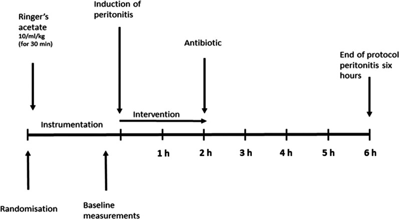
Sepsis is a condition with high morbidity and mortality. Prompt recognition and initiation of treatment is essential. Despite forming an integral part of sepsis management, fluid resuscitation may also lead to volume overload, which in turn is associated with increased mortality. The optimal fluid strategy in sepsis resuscitation is yet to be defined. Hyaluronan, an endogenous glycosaminoglycan with high affinity to water is an important constituent of the endothelial glycocalyx. We hypothesized that exogenously administered hyaluronan would counteract intravascular volume depletion and contribute to endothelial glycocalyx integrity in a fluid restrictive model of peritonitis. In a prospective, blinded model of porcine peritonitis sepsis, we randomized animals to intervention with hyaluronan (n = 8) or 0.9% saline (n = 8). The animals received an infusion of 0.1% hyaluronan 6 ml/kg/h, or the same volume of saline, during the first 2 h of peritonitis. Stroke volume variation and hemoconcentration were comparable in the two groups throughout the experiment. Cardiac output was higher in the intervention group during the infusion of hyaluronan (3.2 ± 0.5 l/min in intervention group vs 2.7 ± 0.2 l/min in the control group) (p = 0.039). The increase in lactate was more pronounced in the intervention group (3.2 ± 1.0 mmol/l in the intervention group and 1.7 ± 0.7 mmol/l in the control group) at the end of the experiment (p < 0.001). Concentrations of surrogate markers of glycocalyx damage; syndecan 1 (0.6 ± 0.2 ng/ml vs 0.5 ± 0.2 ng/ml, p = 0.292), heparan sulphate (1.23 ± 0.2 vs 1.4 ± 0.3 ng/ml, p = 0.211) and vascular adhesion protein 1 (7.0 ± 4.1 vs 8.2 ± 2.3 ng/ml, p = 0.492) were comparable in the two groups at the end of the experiment. In conclusion, hyaluronan did not counteract intravascular volume depletion in early peritonitis sepsis. However, this finding is hampered by the short observation period and a beneficial effect of HMW-HA in peritonitis sepsis cannot be discarded based on the results of the present study.
Keywords: Animal model; Colloid; Fluid therapy; Glycocalyx; Inflammation.
© 2023. European Society of Intensive Care Medicine and Springer Nature Switzerland AG.
Conflict of interest statement
The authors declare that they have no competing interests.
Figures


Similar articles
-
Intravenous fluid resuscitation is associated with septic endothelial glycocalyx degradation.Crit Care. 2019 Jul 23;23(1):259. doi: 10.1186/s13054-019-2534-2. Crit Care. 2019. PMID: 31337421 Free PMC article.
-
Effect of fluid resuscitation on mortality and organ function in experimental sepsis models.Crit Care. 2009;13(6):R186. doi: 10.1186/cc8179. Epub 2009 Nov 23. Crit Care. 2009. PMID: 19930656 Free PMC article.
-
[Dose and timing of normal saline resuscitation on endothelial glycocalyx in early septic shock].Zhonghua Wei Zhong Bing Ji Jiu Yi Xue. 2018 Jul;30(7):629-634. doi: 10.3760/cma.j.issn.2095-4352.2018.07.003. Zhonghua Wei Zhong Bing Ji Jiu Yi Xue. 2018. PMID: 30045788 Chinese.
-
The glycocalyx: a novel diagnostic and therapeutic target in sepsis.Crit Care. 2019 Jan 17;23(1):16. doi: 10.1186/s13054-018-2292-6. Crit Care. 2019. PMID: 30654825 Free PMC article. Review.
-
Surviving sepsis campaign: international guidelines for management of severe sepsis and septic shock: 2012.Crit Care Med. 2013 Feb;41(2):580-637. doi: 10.1097/CCM.0b013e31827e83af. Crit Care Med. 2013. PMID: 23353941
Cited by
-
Managing sepsis and septic shock in an endothelial glycocalyx-friendly way: from the viewpoint of surviving sepsis campaign guidelines.Ann Intensive Care. 2024 Apr 24;14(1):64. doi: 10.1186/s13613-024-01301-6. Ann Intensive Care. 2024. PMID: 38658435 Free PMC article. Review.
-
Role of the endothelial cell glycocalyx in sepsis-induced acute kidney injury.Front Med (Lausanne). 2025 Apr 4;12:1535673. doi: 10.3389/fmed.2025.1535673. eCollection 2025. Front Med (Lausanne). 2025. PMID: 40255592 Free PMC article. Review.
References
LinkOut - more resources
Full Text Sources
Research Materials
