

Incidentally Diagnosed Extraluminal Leiomyosarcoma of Infrarenal Inferior Vena Cava: A Case Report and Literature Review from a Radiologist's Perspective
- PMID: 37733410
- PMCID: PMC9799014
- DOI: 10.15388/Amed.2022.29.2.12
Incidentally Diagnosed Extraluminal Leiomyosarcoma of Infrarenal Inferior Vena Cava: A Case Report and Literature Review from a Radiologist's Perspective
Abstract
Background: Vascular leiomyosarcoma is a rare but most common vascular tumor of the inferior vena cava.
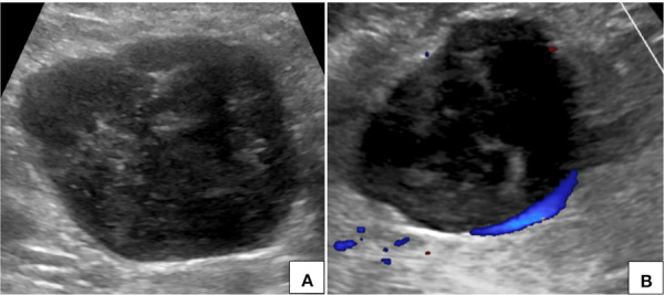
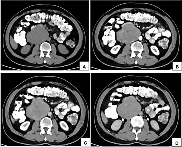
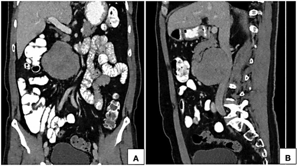
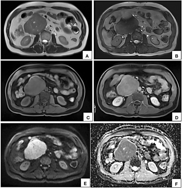
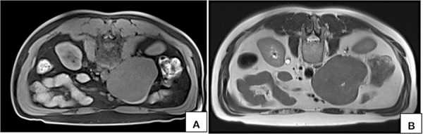
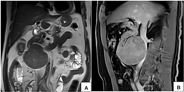
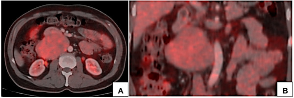
Case presentation: We present the case of an incidentally diagnosed extraluminal leiomyosarcoma of the inferior vena cava in a 62 year old patient who presented with abdominal pain following blunt trauma. Ultrasonography showed a lobulated hypoechoic lesion in the upper abdomen. Computed tomography (CT) and magnetic resonance imaging (MRI) showed a circumscribed lobulated near homogeneously enhancing retroperitoneal lesion in anterior relation to the infrarenal inferior vena cava, right paramedian in location with imperceptible vena caval lumen at the site of maximum contact. In positron emission tomography (PET) CT the lesion showed mild fluorodeoxyglucose (FDG) uptake with no distant metastases. CT guided biopsy with immunohistochemical analysis showed leiomyosarcoma. Patient underwent surgical resection with inferior vena cava reconstruction.
Conclusions: Leiomyosarcoma of the inferior vena cava is a rare tumor of vascular origin. Imaging plays an imperative role in the diagnosis and preoperative evaluation. This article also provides a comprehensive literature review of the radiological features of inferior vena caval leiomyosarcoma that would aid in optimal preoperative characterization and evaluation.
Įvadas: Kraujagyslių lejomiosarkoma yra retas, tačiau dažniausiai pasitaikantis apatinės tuščiosios venos navikas.
Atvejo aprašymas: 62 metų pacientui, kuris kreipėsi į gydymo įstaigą dėl pilvo srities skausmo po neaštriu daiktu sukeltos traumos, buvo atsitiktinai nustatyta neluminalinės infrarenalinės apatinės tuščiosios venos lejomiosarkoma.Ultragarso tyrimas atskleidė skiltinį hipoechogenišką pažeidimą viršutinėje pilvo srityje. Kompiuterinė tomografija (KT) ir magnetinio rezonanso tomografija (MRT) parodė, kad priešakinėje infrarenalinėje apatinėje tuščiojoje venoje yra apibrėžtas skiltinis, beveik homogeniškai intensyvėjantis retroperitoninis pažeidimas, kuris didžiausio sąlyčio vietoje su tuščiosios venos spindžiu į dešinę nuo centro yra beveik nematomas. Pozitronų emisijos tomografijos (PET) metu pažeidimas parodė nedidelį fluorodeoksigliukozės (FDG) įsisavinimą be jokių tolimų metastazių. Atlikus kompiuterine tomografija paremta biopsiją su imunohistochemine analize, nustatyta lejomiosarkoma. Pacientui buvo atlikta chirurginė rezekcija ir apatinės tuščiosios venos rekonstrukcija.
Išvados: Apatinės tuščiosios venos lejomiosarkoma yra retas kraujagyslių sistemos auglys. Diagnozei ir vertinimui iki operacijos būtina peržiūrėti vaizdinę medžiagą. Šiame straipsnyje taip pat pateikiama išsami literatūros apžvalga apie radiologines apatinės tuščiosios venos lejomiosarkomos savybes, kurios pagerintų optimalų priešoperacinį įvertinimą.
Keywords: Leiomyosarcoma; imaging features; inferior vena cava; retroperitoneal tumor.
Copyright © 2022 Ebinesh A, Aanchal Ashta, Satyam, Gaurav Shanker Pradhan, Rohin Sharma, Prince Das. Published by Vilnius University Press.
Conflict of interest statement
The Autrhors confirm that this work is original and has been published only in a pre-print service (www.researchsquare.com), the DOI is the following: https://doi.org/10.21203/rs.3.rs-1918169/v1.
Figures
Similar articles
-
Cine magnetic resonance imaging in evaluating retroperitoneal leiomyosarcoma arising from the inferior vena cava.IJU Case Rep. 2023 Oct 27;7(1):30-33. doi: 10.1002/iju5.12660. eCollection 2024 Jan. IJU Case Rep. 2023. PMID: 38173447 Free PMC article.
-
Infrarenal Vena Cava Leiomyosarcoma Treated With Surgical Resection and Vascular Reconstruction.Cureus. 2021 Jun 21;13(6):e15808. doi: 10.7759/cureus.15808. eCollection 2021 Jun. Cureus. 2021. PMID: 34306875 Free PMC article.
-
Primary leiomyosarcoma of the inferior vena cava in a pediatric case: a case report and literature review.Surg Case Rep. 2023 Apr 6;9(1):52. doi: 10.1186/s40792-023-01630-x. Surg Case Rep. 2023. PMID: 37022631 Free PMC article.
-
Leiomyosarcoma of the inferior vena cava: analysis and search of world literature on 141 patients and report of three new cases.J Vasc Surg. 1991 Nov;14(5):688-99. doi: 10.1067/mva.1991.30426. J Vasc Surg. 1991. PMID: 1942380 Review.
-
Leiomyosarcoma of the inferior vena cava: a case report and review of literature.Int Surg. 2005 Nov-Dec;90(5):262-5. Int Surg. 2005. PMID: 16625943 Review.
References
-
- Perl L. Ein fall von sarkom der vena cava inferior. Virchows Arch. 1871;53:378–383.
-
- Bower TC, Stanson A. Diagnosis and management of tumors of the inferior vena cava. In: Rutherford RB, ed. Vascular surgery. WB Saunders; 2000:2077–2092.
Publication types
LinkOut - more resources
Full Text Sources