

Astragalus polysaccharide inhibits the development of urothelial carcinoma by activating AMPK signaling to induce BENC1-xCT complex formation
- PMID: 37733667
- PMCID: PMC10564440
- DOI: 10.18632/aging.205007
Astragalus polysaccharide inhibits the development of urothelial carcinoma by activating AMPK signaling to induce BENC1-xCT complex formation
Abstract
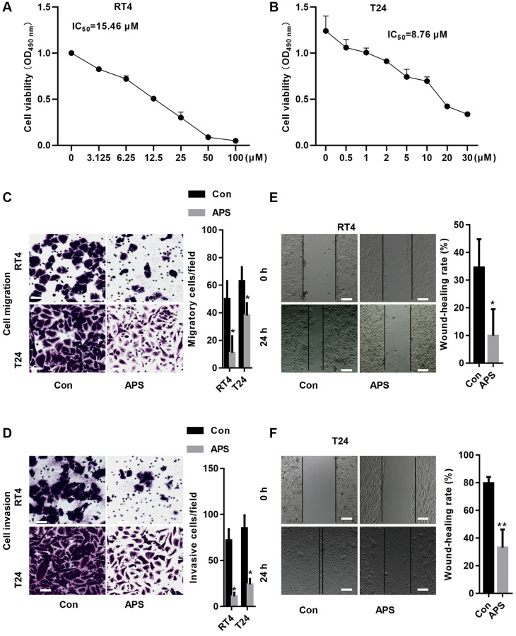
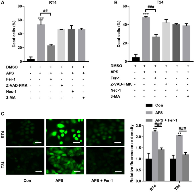
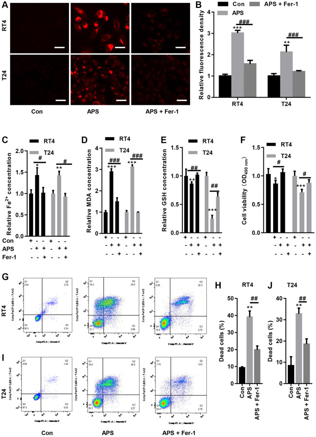
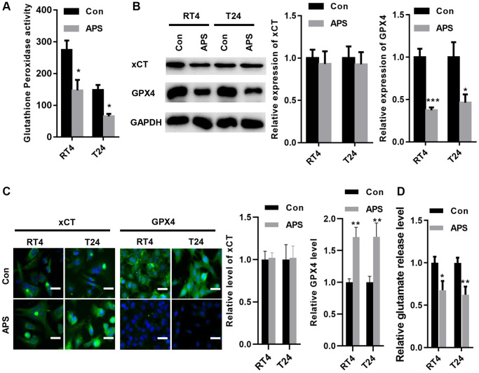
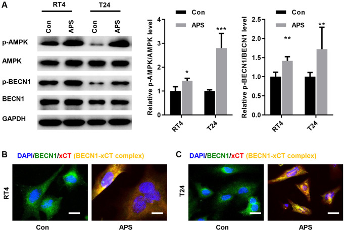
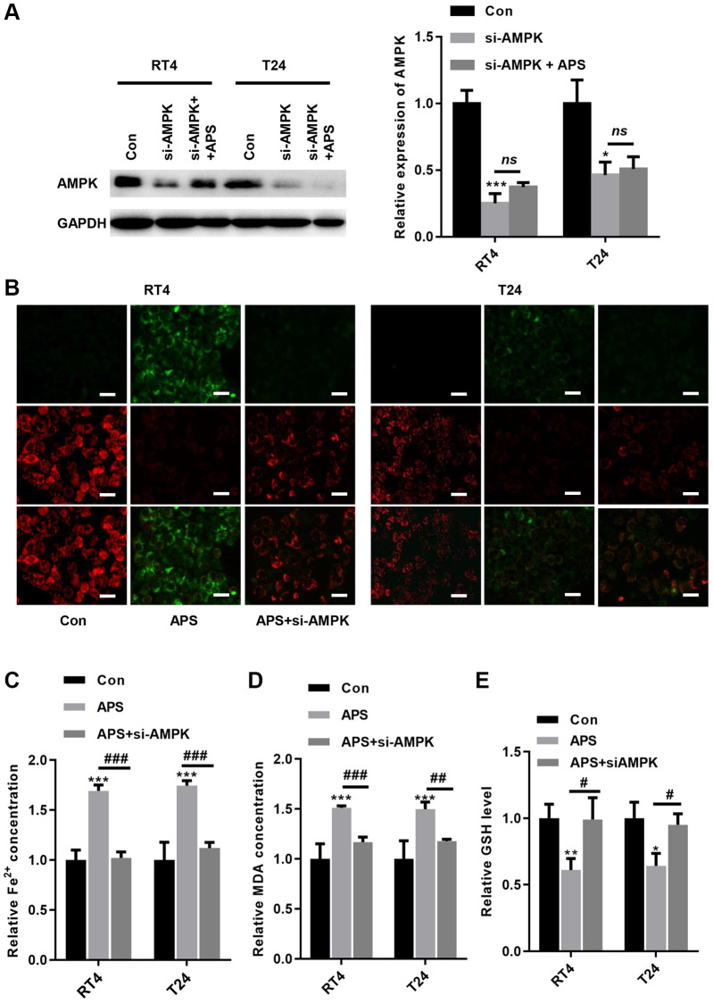
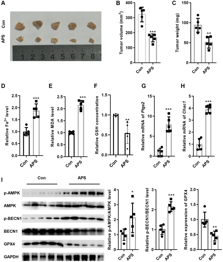
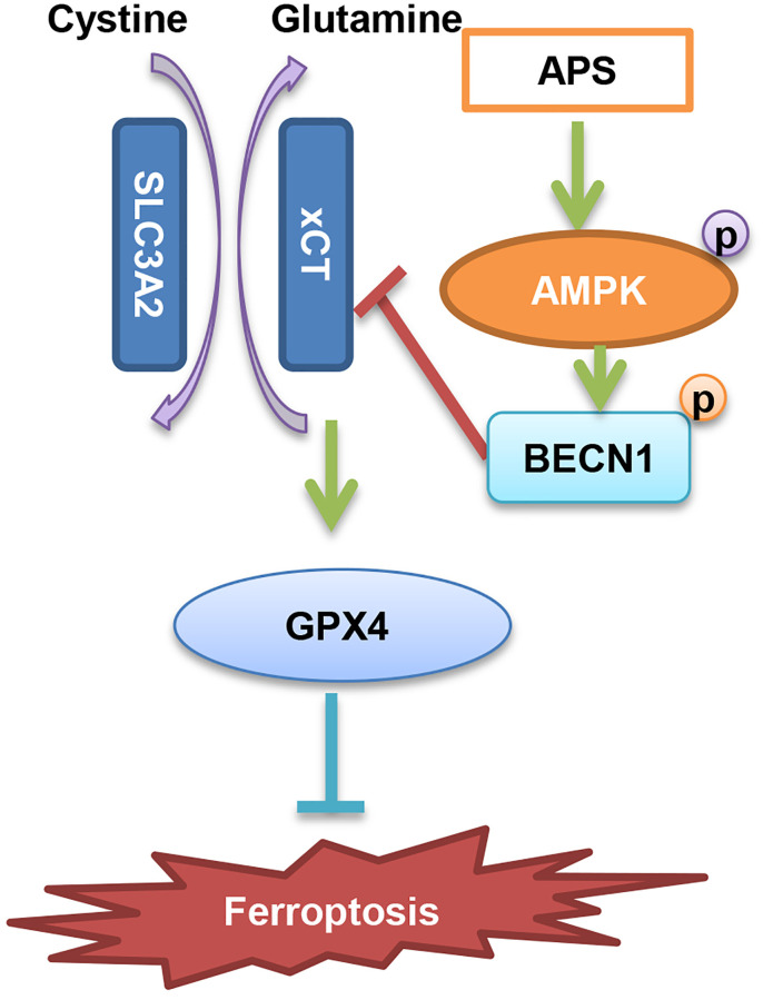
In recent years, the incidence of urothelial carcinoma (UC) has been high in men. The aim of this study was to investigate whether astragalus polysaccharide (APS) could inhibit the development of UC and the specific molecular mechanism. Our data showed that APS inhibited the proliferation of UC cells in a dose-dependent manner, and APS reduced the migratory capacity of RT4 and T24 cells. Further studies revealed that the ferroptosis inhibitor ferrostatin-1 (Fer-1) reversed APS-induced cell death, intracellular Fe2+ and malondialdehyde (MDA) accumulation, and lipid peroxidation product deposition. The Western blot and immunofluorescence results showed that APS significantly inhibited the expression of glutathione peroxidase 4 (GPX4) but did not alter the protein level of solute carrier family 7 member 11 (xCT, SLC7A11). Further analysis revealed that APS reduced the activity of xCT in RT4 and T24 cells. Moreover, APS significantly increased the phosphorylation levels of protein kinase AMP-activated catalytic subunit alpha 1 (AMPK) and BECN1 in RT4 and T24 cells, which induced the formation of the BECN1-xCT complex. However, when AMPK was silenced in RT4 and T24 cells, APS-induced ferroptosis was reversed to some extent, indicating that APS-mediated ferroptosis involves AMPK signaling. Moreover, APS has been shown to inhibit tumor growth in nude mice in vivo. In summary, our study demonstrated for the first time that APS could promote the formation of the BECN1-xCT complex in UC cells by activating AMPK/BECN1 signaling, which inhibited the activity of xCT to reduce GPX4 expression, thereby inducing ferroptosis and ultimately inhibiting UC progression.
Keywords: AMPK activation; BECN1-xCT complex; astragalus polysaccharides; ferroptosis; urothelial carcinoma.
Conflict of interest statement
Figures
Similar articles
-
Capsaicin induces ferroptosis via suppression of SLC7A11 activity and upregulation of ACSL4 mediated by AMPK in tongue squamous cell carcinoma.Front Oncol. 2025 May 14;15:1532555. doi: 10.3389/fonc.2025.1532555. eCollection 2025. Front Oncol. 2025. PMID: 40438695 Free PMC article.
-
AMPK-Mediated BECN1 Phosphorylation Promotes Ferroptosis by Directly Blocking System Xc- Activity.Curr Biol. 2018 Aug 6;28(15):2388-2399.e5. doi: 10.1016/j.cub.2018.05.094. Epub 2018 Jul 26. Curr Biol. 2018. PMID: 30057310 Free PMC article.
-
SIRT3-mediated autophagy contributes to ferroptosis-induced anticancer by inducing the formation of BECN1-SLC7A11 complex.Biochem Pharmacol. 2023 Jul;213:115592. doi: 10.1016/j.bcp.2023.115592. Epub 2023 May 16. Biochem Pharmacol. 2023. PMID: 37196680
-
A novel anticancer property of Lycium barbarum polysaccharide in triggering ferroptosis of breast cancer cells.J Zhejiang Univ Sci B. 2022 Apr 15;23(4):286-299. doi: 10.1631/jzus.B2100748. J Zhejiang Univ Sci B. 2022. PMID: 35403384 Free PMC article. English.
-
Astragalus polysaccharide stimulates glucose uptake in L6 myotubes through AMPK activation and AS160/TBC1D4 phosphorylation.Acta Pharmacol Sin. 2013 Jan;34(1):137-45. doi: 10.1038/aps.2012.133. Epub 2012 Oct 29. Acta Pharmacol Sin. 2013. PMID: 23103623 Free PMC article.
Cited by
-
Jianpi Yangzheng Xiaozheng granule induced ferroptosis to suppress gastric cancer progression through reprogramming lipid metabolism via SCD1/Wnt/β-catenin axis.Front Mol Biosci. 2025 Feb 25;12:1523494. doi: 10.3389/fmolb.2025.1523494. eCollection 2025. Front Mol Biosci. 2025. PMID: 40070686 Free PMC article.
-
Capsaicin induces ferroptosis via suppression of SLC7A11 activity and upregulation of ACSL4 mediated by AMPK in tongue squamous cell carcinoma.Front Oncol. 2025 May 14;15:1532555. doi: 10.3389/fonc.2025.1532555. eCollection 2025. Front Oncol. 2025. PMID: 40438695 Free PMC article.
-
Advances in research on the anti-tumor mechanism of Astragalus polysaccharides.Front Oncol. 2024 Mar 6;14:1334915. doi: 10.3389/fonc.2024.1334915. eCollection 2024. Front Oncol. 2024. PMID: 38515577 Free PMC article. Review.
-
Combination of astragalus polysaccharide with Diosbulbin B exerts an enhanced antitumor effect in BRAFmut papillary thyroid cancer with decreased liver toxicity.Cancer Cell Int. 2025 Jul 2;25(1):245. doi: 10.1186/s12935-025-03853-4. Cancer Cell Int. 2025. PMID: 40604967 Free PMC article.
References
-
- DeGeorge KC, Holt HR, Hodges SC. Bladder Cancer: Diagnosis and Treatment. Am Fam Physician. 2017; 96:507–14. - PubMed
Publication types
MeSH terms
Substances
LinkOut - more resources
Full Text Sources
Medical
Miscellaneous
